Copyright
©The Author(s) 2023.
World J Clin Cases. Feb 26, 2023; 11(6): 1410-1418
Published online Feb 26, 2023. doi: 10.12998/wjcc.v11.i6.1410
Published online Feb 26, 2023. doi: 10.12998/wjcc.v11.i6.1410
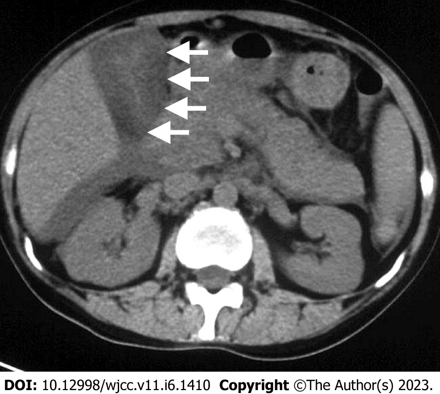
Figure 1 Computed tomography scan of the gallbladder and its surroundings.
Axial computed tomography image confirmed distended gallbladder (9.1 cm × 4.1 cm) with an evenly thickened, hydropic gallbladder wall (approximately 1.8 cm). Pericholecystic and hepatic fluid was also seen. No calculi were present.
- Citation: Chang CH, Wang YY, Jiao Y. Hepatitis A virus-associated acute acalculous cholecystitis in an adult-onset Still’s disease patient: A case report and review of the literature. World J Clin Cases 2023; 11(6): 1410-1418
- URL: https://www.wjgnet.com/2307-8960/full/v11/i6/1410.htm
- DOI: https://dx.doi.org/10.12998/wjcc.v11.i6.1410


