Copyright
©The Author(s) 2023.
World J Clin Cases. Feb 26, 2023; 11(6): 1379-1384
Published online Feb 26, 2023. doi: 10.12998/wjcc.v11.i6.1379
Published online Feb 26, 2023. doi: 10.12998/wjcc.v11.i6.1379
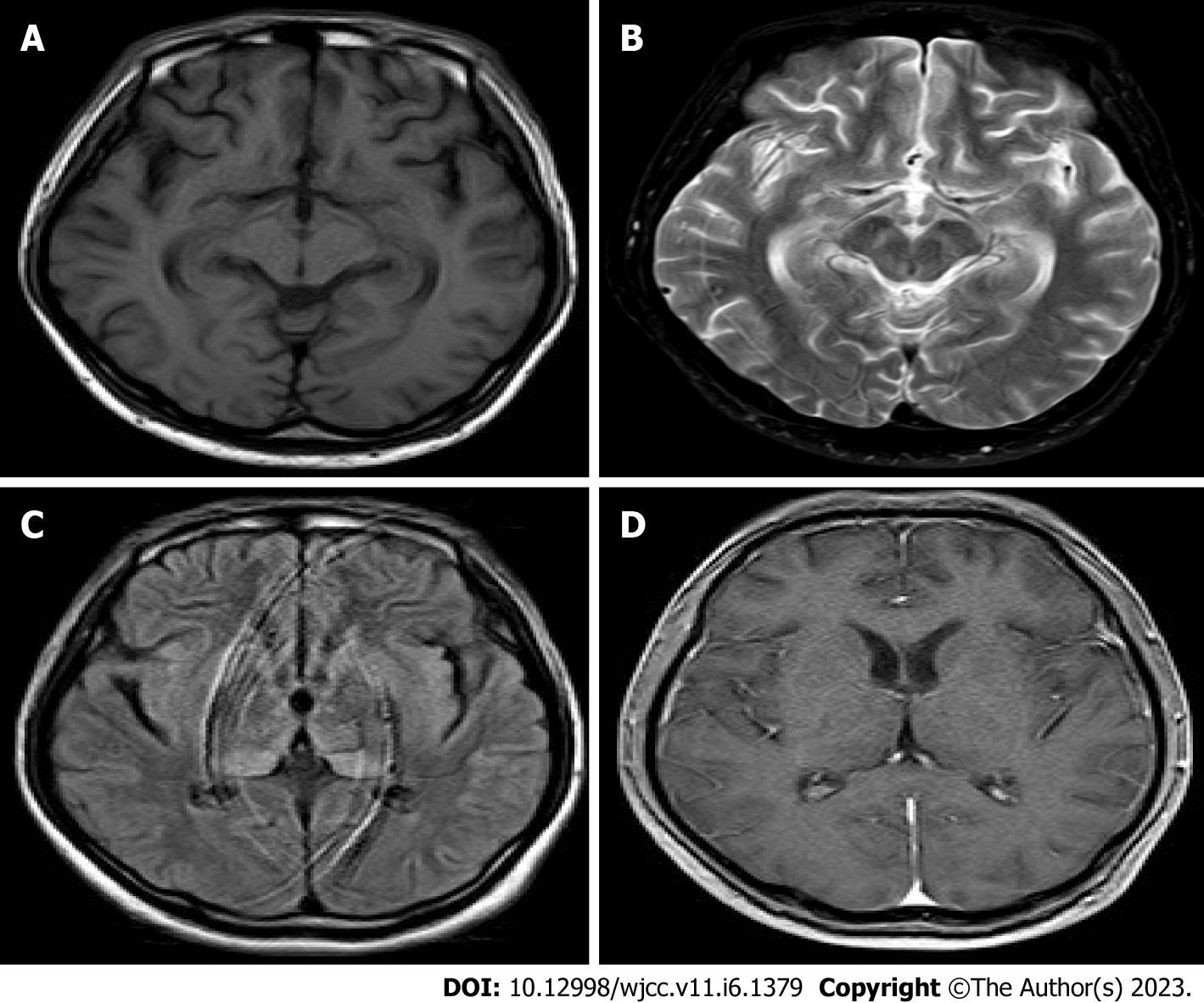
Figure 2 Head magnetic resonance image.
A: T1 image: No obvious abnormalities; B: T2 image: Bilateral medial temporal lobe high signal; C: T2 Flair image: High signal in both basal ganglia and thalamus; D: T1 enhanced image.
- Citation: Huang P. Epidemic Japanese B encephalitis combined with contactin-associated protein-like 2 antibody-positive autoimmune encephalitis: A case report. World J Clin Cases 2023; 11(6): 1379-1384
- URL: https://www.wjgnet.com/2307-8960/full/v11/i6/1379.htm
- DOI: https://dx.doi.org/10.12998/wjcc.v11.i6.1379


