Copyright
©The Author(s) 2023.
World J Clin Cases. Feb 26, 2023; 11(6): 1356-1364
Published online Feb 26, 2023. doi: 10.12998/wjcc.v11.i6.1356
Published online Feb 26, 2023. doi: 10.12998/wjcc.v11.i6.1356
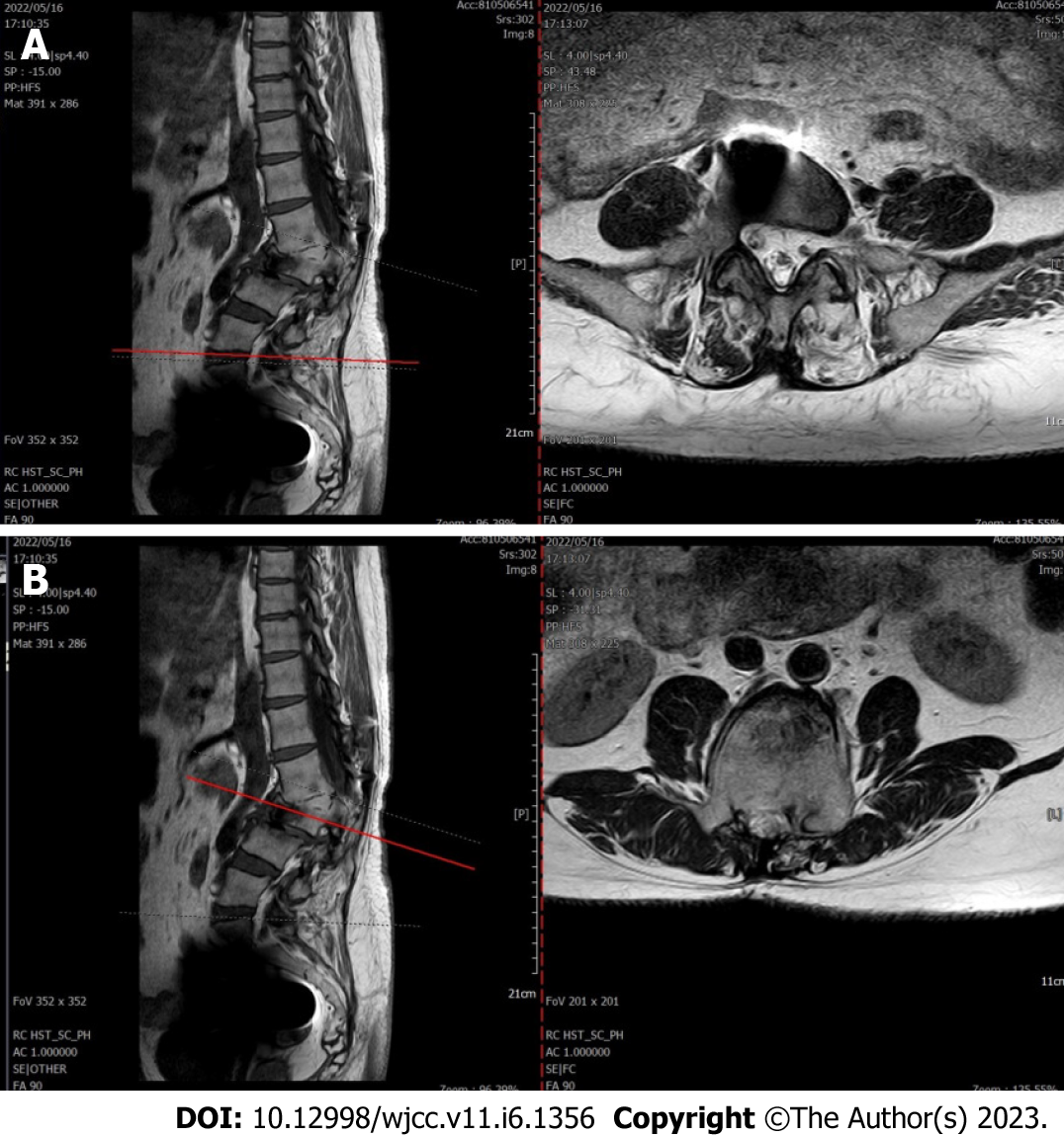
Figure 5 The lumbar spine magnetic resonance imaging.
Lumbar vertebrae protrude backward, L3 vertebrae show wedge-shaped changes, spinal canal stenosis on the same level of L3, intervertebral space narrowing on the same level of L2-3, endplate inflammation in the L3-4 intervertebral space, Schmorl node formation near T10-11 vertebral body, degeneration and bulge of L3-4, L4-5 and L5-S1 intervertebral discs, and many abnormal strip signals in the filum terminale. A: L5-S1 Plain magnetic resonance imaging (MRI) scan of L5-S1 intervertebral disc; B: L3 MRI plain scan.
- Citation: Liu YD, Deng Q, Li JJ, Yang HY, Han XF, Zhang KD, Peng RD, Xiang QQ. Post-traumatic cauda equina nerve calcification: A case report. World J Clin Cases 2023; 11(6): 1356-1364
- URL: https://www.wjgnet.com/2307-8960/full/v11/i6/1356.htm
- DOI: https://dx.doi.org/10.12998/wjcc.v11.i6.1356


