Copyright
©The Author(s) 2023.
World J Clin Cases. Feb 26, 2023; 11(6): 1356-1364
Published online Feb 26, 2023. doi: 10.12998/wjcc.v11.i6.1356
Published online Feb 26, 2023. doi: 10.12998/wjcc.v11.i6.1356
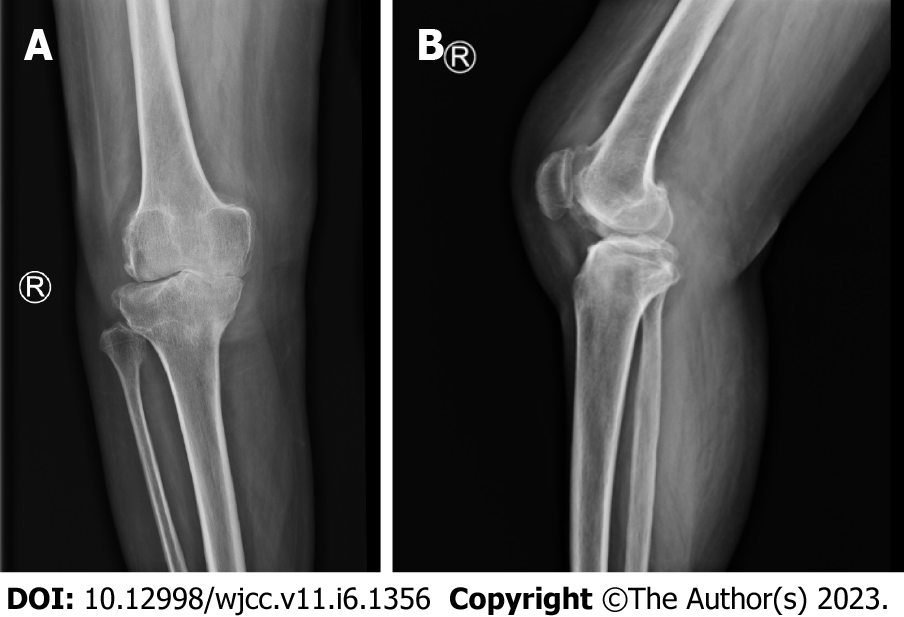
Figure 3 The X-ray film of the right knee joint.
The right knee joint space was narrow inside and wide outside, the articular surface was sclerotic, and the lower edge of the articular surface was spotted with low density. Hyperosteogeny appeared on the upper edge of patella, tibial plateau, tibial intercondylar crest and femoral condyle. The density of the suprapatellar bursa is increased and the surrounding soft tissue is swollen. A: Orthostatic Knee joint X-ray film; B: Lateral Knee joint X-ray film.
- Citation: Liu YD, Deng Q, Li JJ, Yang HY, Han XF, Zhang KD, Peng RD, Xiang QQ. Post-traumatic cauda equina nerve calcification: A case report. World J Clin Cases 2023; 11(6): 1356-1364
- URL: https://www.wjgnet.com/2307-8960/full/v11/i6/1356.htm
- DOI: https://dx.doi.org/10.12998/wjcc.v11.i6.1356


