Copyright
©The Author(s) 2023.
World J Clin Cases. Feb 16, 2023; 11(5): 1217-1223
Published online Feb 16, 2023. doi: 10.12998/wjcc.v11.i5.1217
Published online Feb 16, 2023. doi: 10.12998/wjcc.v11.i5.1217
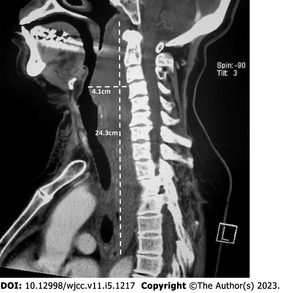
Figure 2 Preoperative sagittal venous phase contrast-enhanced computed tomography image of the neck (median plane).
The soft tissue density lesion extends from the neck to the mediastinum and involves the posterior pharyngeal wall, middle and upper oesophagus, posterior cervical trachea, and thoracic trachea. The nasopharynx, oropharynx, and laryngeal pharynx are compressed and narrowed. The trachea is compressed.
- Citation: Han YZ, Zhou Y, Peng Y, Zeng J, Zhao YQ, Gao XR, Zeng H, Guo XY, Li ZQ. Difficult airway due to cervical haemorrhage caused by spontaneous rupture of a parathyroid adenoma: A case report. World J Clin Cases 2023; 11(5): 1217-1223
- URL: https://www.wjgnet.com/2307-8960/full/v11/i5/1217.htm
- DOI: https://dx.doi.org/10.12998/wjcc.v11.i5.1217


