Copyright
©The Author(s) 2023.
World J Clin Cases. Feb 6, 2023; 11(4): 962-971
Published online Feb 6, 2023. doi: 10.12998/wjcc.v11.i4.962
Published online Feb 6, 2023. doi: 10.12998/wjcc.v11.i4.962
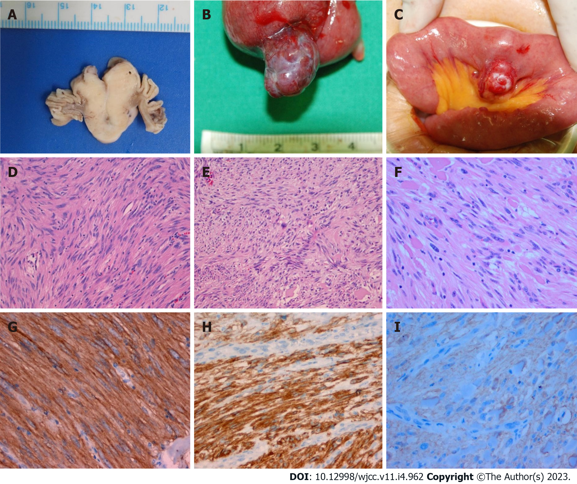
Figure 8 Macroscopic and microscopic examination.
A: Macroscopic cross section shows a gray-white solid tumor; B: Macroscopic examination shows a 2 cm (longest diameter) encapsulated lobulating mass; C: Macroscopic examination shows a 2.5 cm sized encapsulated round mass; D-F: Microscopic examinations show that the tumors are composed of spindle cells having high cellularity (hematoxylin-eosin staining, × 100); G-I: Immunohistochemically, these tumors are positive for CD117 (c-kit stain, × 200). (A, D, and G: Patient 1; B, E, and H: Patient 2; C, F, and I: Patient 3).
- Citation: Lee J, Kim S, Kim D, Lee S, Ryu K. Three cases of jejunal tumors detected by standard upper gastrointestinal endoscopy: A case series. World J Clin Cases 2023; 11(4): 962-971
- URL: https://www.wjgnet.com/2307-8960/full/v11/i4/962.htm
- DOI: https://dx.doi.org/10.12998/wjcc.v11.i4.962


