Copyright
©The Author(s) 2023.
World J Clin Cases. Feb 6, 2023; 11(4): 931-937
Published online Feb 6, 2023. doi: 10.12998/wjcc.v11.i4.931
Published online Feb 6, 2023. doi: 10.12998/wjcc.v11.i4.931
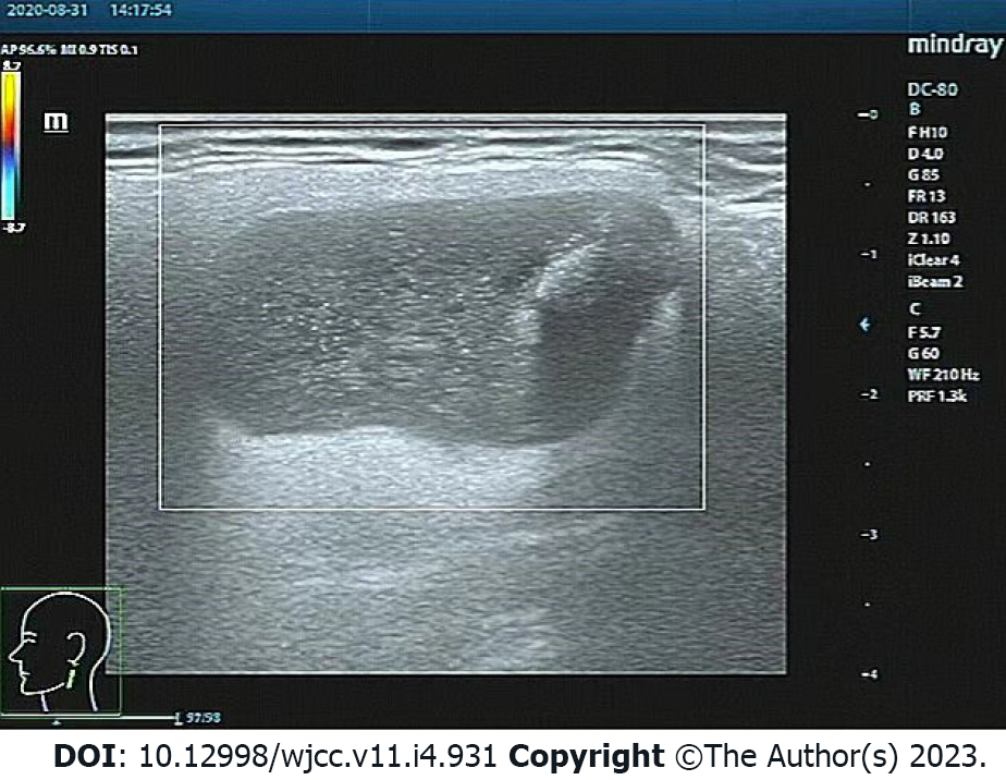
Figure 1 Color ultrasound of the neck.
It reveals a 2.2 cm × 2.2 cm hypoechoic mass in the left parotid gland, with a clear boundary, coarse calcification foci, and no blood flow signal.
- Citation: Liao Y, Li YJ, Hu XW, Wen R, Wang P. Benign lymphoepithelial cyst of parotid gland without human immunodeficiency virus infection: A case report . World J Clin Cases 2023; 11(4): 931-937
- URL: https://www.wjgnet.com/2307-8960/full/v11/i4/931.htm
- DOI: https://dx.doi.org/10.12998/wjcc.v11.i4.931


