Copyright
©The Author(s) 2023.
World J Clin Cases. Feb 6, 2023; 11(4): 922-930
Published online Feb 6, 2023. doi: 10.12998/wjcc.v11.i4.922
Published online Feb 6, 2023. doi: 10.12998/wjcc.v11.i4.922
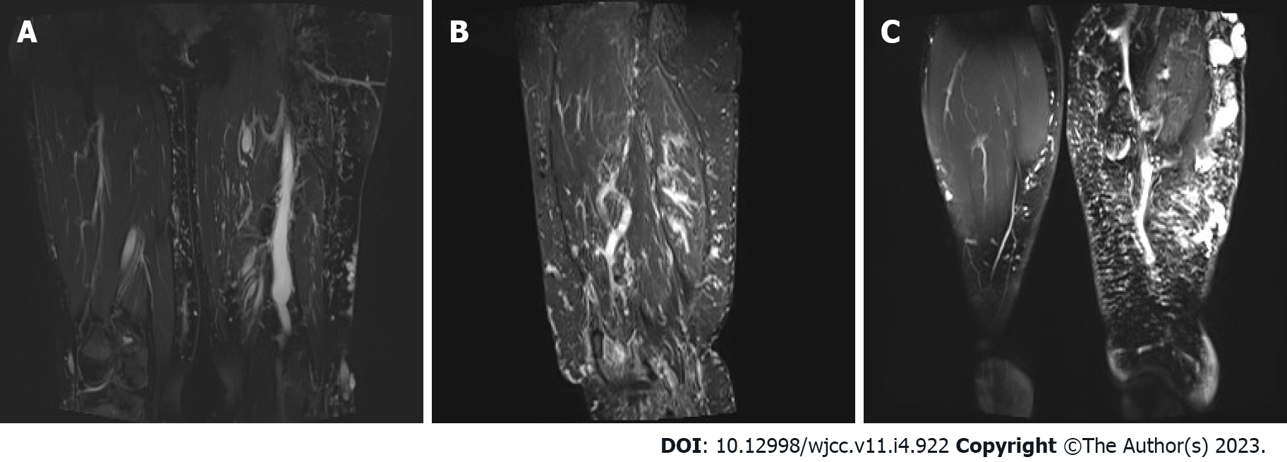
Figure 3 Left lower extremity magnetic resonance imaging plain scan + enhancement.
A-C: Obvious enlargement, swelling, and extensive reticular signal abnormality can be seen in the left lower extremity (Lymphadenopathy). The subcutaneous varicose veins can be noted on the left leg.
- Citation: Li LL, Xie R, Li FQ, Huang C, Tuo BG, Wu HC. Easily misdiagnosed complex Klippel-Trenaunay syndrome: A case report. World J Clin Cases 2023; 11(4): 922-930
- URL: https://www.wjgnet.com/2307-8960/full/v11/i4/922.htm
- DOI: https://dx.doi.org/10.12998/wjcc.v11.i4.922


