Copyright
©The Author(s) 2023.
World J Clin Cases. Feb 6, 2023; 11(4): 756-763
Published online Feb 6, 2023. doi: 10.12998/wjcc.v11.i4.756
Published online Feb 6, 2023. doi: 10.12998/wjcc.v11.i4.756
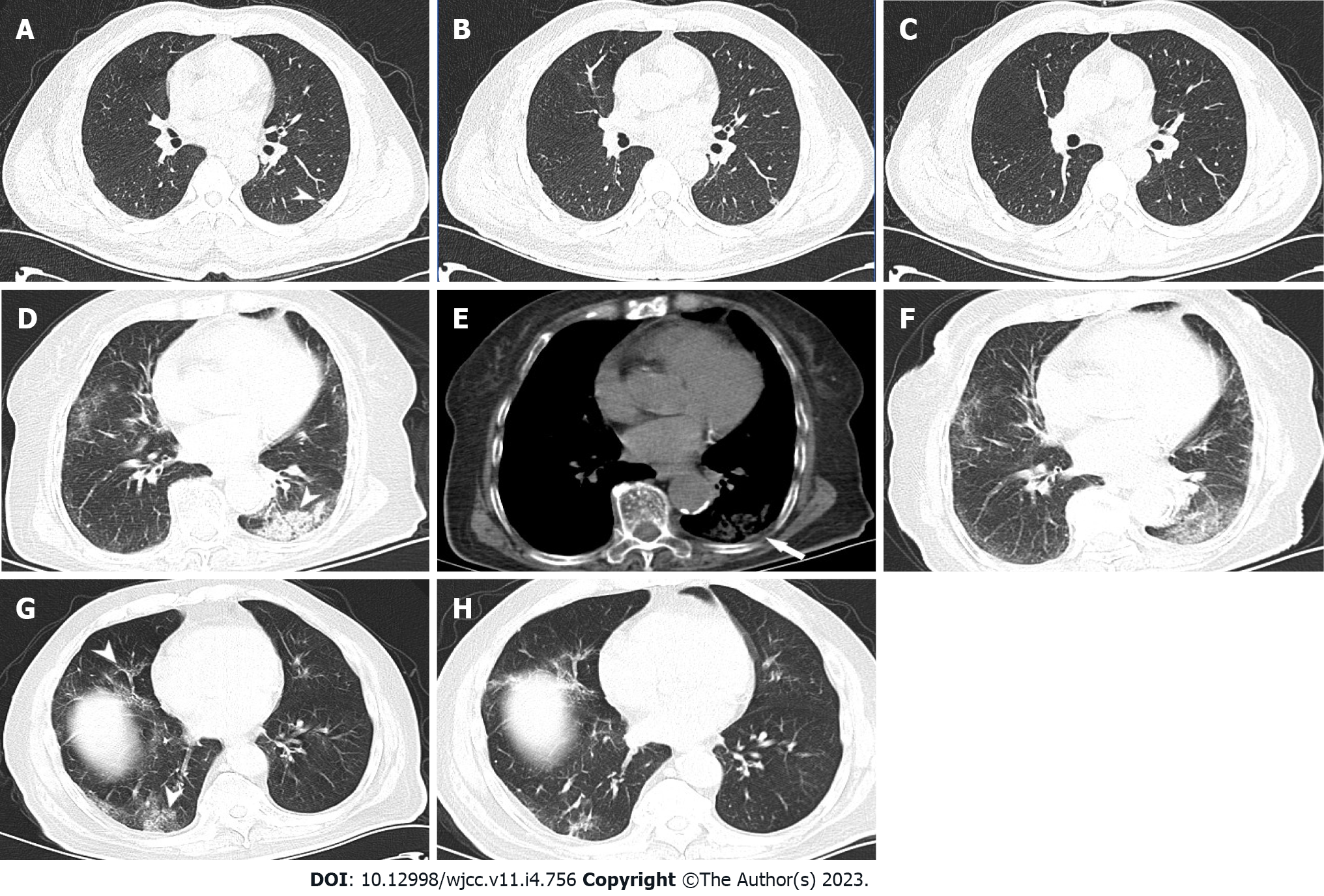
Figure 1 Computed tomography.
A and B: A 48-year-old man had a history of close contact with patients mildly infected with SARS-CoV-2 Omicron variant. He had fever and mild cough for 3 d, with nucleic acid polymerase chain reaction positivity [cycle threshold (Ct) value, 35.43], leukocyte count of 7.26 × 10⁹/L, neutrophil proportion of 58.8%, lymphocyte proportion of 32.9%, hypersensitive C-reactive protein content of < 0.5 mg/L (reference range, 0-8), and serum amyloid A (rapid method) content of < 5.0 mg/L (reference value < 10). The pulmonary window of the chest computed tomography (CT) scan revealed a focal high-density infection in the long diameter of the dorsal segment of the left lower lobe (< 2 cm) (A, short arrow); CT re-examination after 4 d revealed a decreased lesion density and slightly increased volume in the dorsal segment of the left lower lobe (B); 1 wk later, CT images showed that most of the lesions were dissipated and absorbed (C); D-F: A 93-year-old woman had a history of close contact with asymptomatic patients infected with SARS-CoV-2 Omicron variant. She had a high fever, cough, and expectoration for 4 d, with nucleic acid polymerase chain reaction positivity (Ct value, 23.97), leukocyte count of 7.41 × 10⁹/L, neutrophil proportion of 63.3%, lymphocyte proportion of 22.25% (close to the lower limit of normal value), hypersensitive C-reactive protein content of 31.48 mg/L↑, serum amyloid A (rapid method) content of > 200 mg/L↑, partial pressure of carbon dioxide of 4.65↓, and D-dimer content of 4.25↑. The pulmonary window of the chest CT scan revealed consolidation shadows in the dorsal segment of the left lower lobe (short arrow) accompanied with a small amount of effusion in the adjacent pleural cavity, and scattered patchy, slightly high-density infection foci in both lungs, suggestive of an infection (D); the mediastinal window of the same CT revealed a small amount of effusion in the left pleural cavity (long arrow) (E); CT re-examination after 5 d revealed a decreased density of the consolidation infection foci in the dorsal segment of the left lower lobe and partial absorption of other infection foci in both lungs (F); G and H: An 81-year-old man had a history of close contact with his wife who had asymptomatic SARS-CoV-2 Omicron infection. He had fever, cough, and expectoration for 4 d, with nucleic acid polymerase chain reaction positivity (Ct value, 26.3), leukocyte count of 7.73 × 10⁹/L, neutrophil proportion of 61.9%, lymphocyte proportion of 20.1% (close to the lower limit of normal value), hypersensitive C-reactive protein content of 7.42 mg/L, and serum amyloid A (rapid method) content of 16.6 mg/L↑. The pulmonary window of the chest CT scan revealed scattered patchy ground-glass density shadows in the right middle and lower lobes (short arrows) (G); CT re-examination after 6 d revealed shrinkage and partial absorption of most of the infected foci in the right lung (H).
- Citation: Ying WF, Chen Q, Jiang ZK, Hao DG, Zhang Y, Han Q. Chest computed tomography findings of the Omicron variants of SARS-CoV-2 with different cycle threshold values. World J Clin Cases 2023; 11(4): 756-763
- URL: https://www.wjgnet.com/2307-8960/full/v11/i4/756.htm
- DOI: https://dx.doi.org/10.12998/wjcc.v11.i4.756


