Copyright
©The Author(s) 2023.
World J Clin Cases. Dec 26, 2023; 11(36): 8527-8534
Published online Dec 26, 2023. doi: 10.12998/wjcc.v11.i36.8527
Published online Dec 26, 2023. doi: 10.12998/wjcc.v11.i36.8527
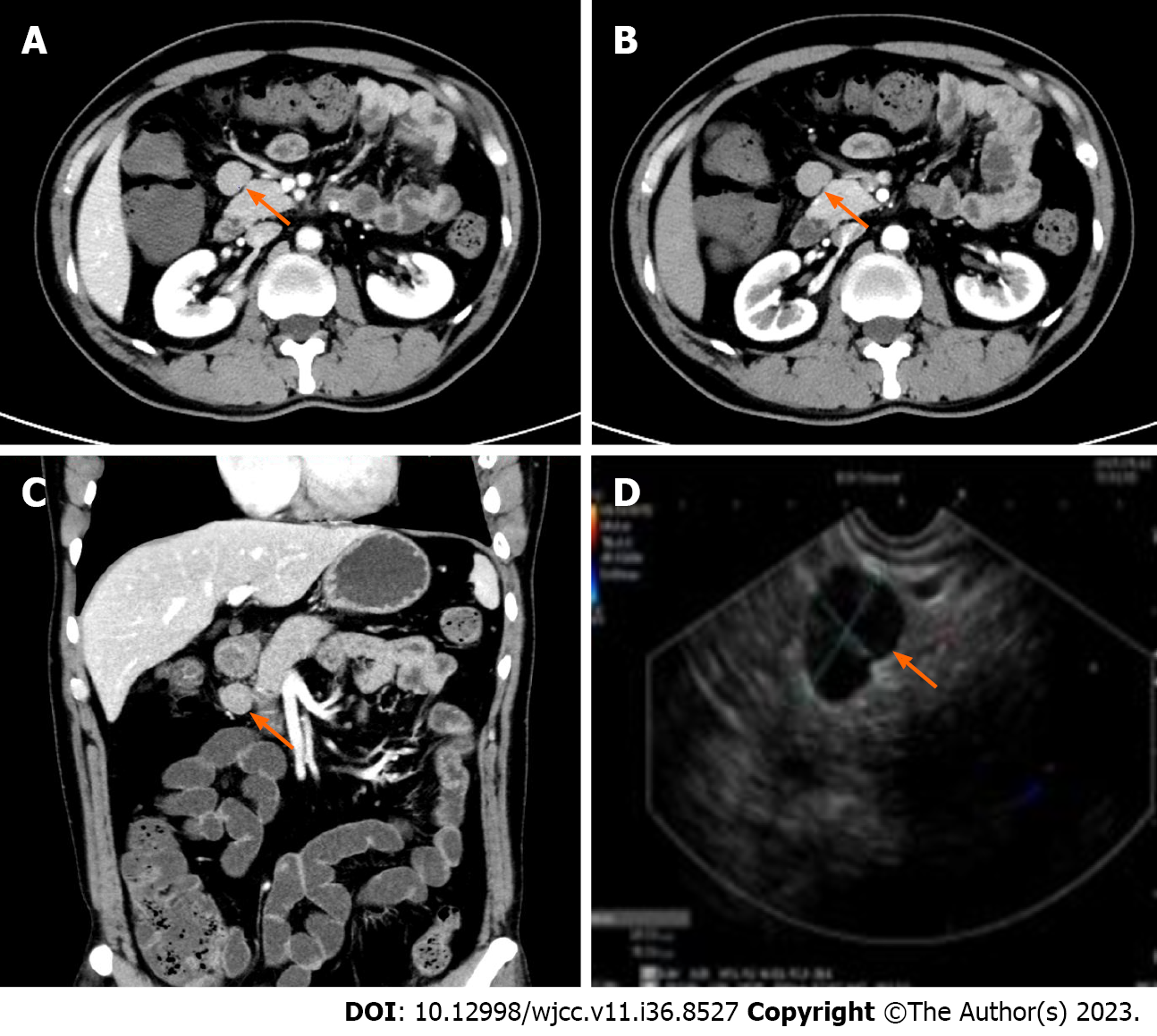
Figure 1 Abdominal computed tomography showed a soft tissue occupying focus in front of the pancreas in Case 1.
The lesion had a quasi-round soft tissue density, the edge was smooth, and the enhanced scan showed obvious uniform and continuous enhancement. A: Venous phase; B: Arterial phase; C: Coronal imaging; D: Ultrasonic gastroscopic image.
- Citation: Gao JW, Shi ZY, Zhu ZB, Xu XR, Chen W. Intraperitoneal hyaline vascular Castleman disease: Three case reports. World J Clin Cases 2023; 11(36): 8527-8534
- URL: https://www.wjgnet.com/2307-8960/full/v11/i36/8527.htm
- DOI: https://dx.doi.org/10.12998/wjcc.v11.i36.8527


