Copyright
©The Author(s) 2023.
World J Clin Cases. Dec 16, 2023; 11(35): 8404-8410
Published online Dec 16, 2023. doi: 10.12998/wjcc.v11.i35.8404
Published online Dec 16, 2023. doi: 10.12998/wjcc.v11.i35.8404
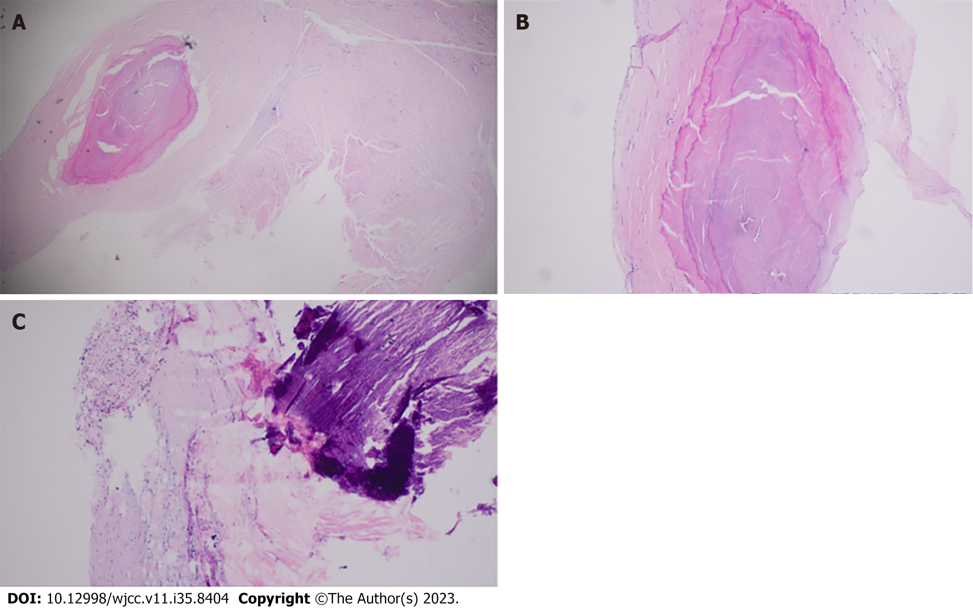
Figure 5 Phlebosclerosis: pathological examination of the vein resembling a wooden rod.
A: The cross-section of the vein during surgery shows venous fibrosis, calcification, and thickening of the vein wall. Extensive collagen deposition is observed on the vein wall, with hyaline degeneration and venous sclerosis causing closure of the venous lumen (× 40 magnification); B: × 100 magnification; C: Calcification in the venous lumen and inflammatory cell infiltration in the venous wall (× 100 magnification).
- Citation: Ren SY, Qian SY, Gao RD. Phlebosclerosis: An overlooked complication of varicose veins that affects clinical outcome: A case report. World J Clin Cases 2023; 11(35): 8404-8410
- URL: https://www.wjgnet.com/2307-8960/full/v11/i35/8404.htm
- DOI: https://dx.doi.org/10.12998/wjcc.v11.i35.8404


