Copyright
©The Author(s) 2023.
World J Clin Cases. Dec 6, 2023; 11(34): 8228-8234
Published online Dec 6, 2023. doi: 10.12998/wjcc.v11.i34.8228
Published online Dec 6, 2023. doi: 10.12998/wjcc.v11.i34.8228
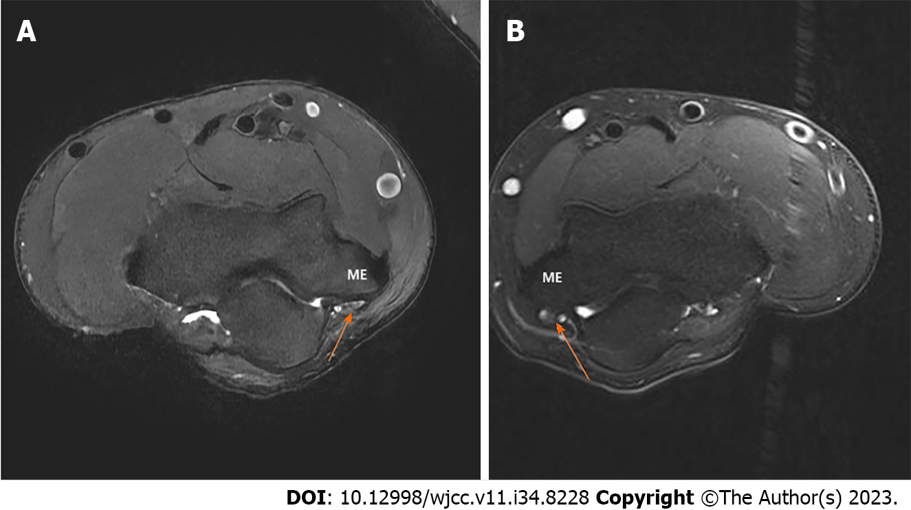
Figure 1 Pre-operative axial T2-weighted magnetic resonance image shows the high signal change and swelling of the ulnar nerve.
A: Rt. Elbow; B: Lt. Elbow. Arrow: Ulnar nerve, ME: Medial epicondyle.
- Citation: Cho CH, Lim KH, Kim DH. Bilateral snapping triceps syndrome: A case report. World J Clin Cases 2023; 11(34): 8228-8234
- URL: https://www.wjgnet.com/2307-8960/full/v11/i34/8228.htm
- DOI: https://dx.doi.org/10.12998/wjcc.v11.i34.8228


