Copyright
©The Author(s) 2023.
World J Clin Cases. Nov 26, 2023; 11(33): 8038-8043
Published online Nov 26, 2023. doi: 10.12998/wjcc.v11.i33.8038
Published online Nov 26, 2023. doi: 10.12998/wjcc.v11.i33.8038
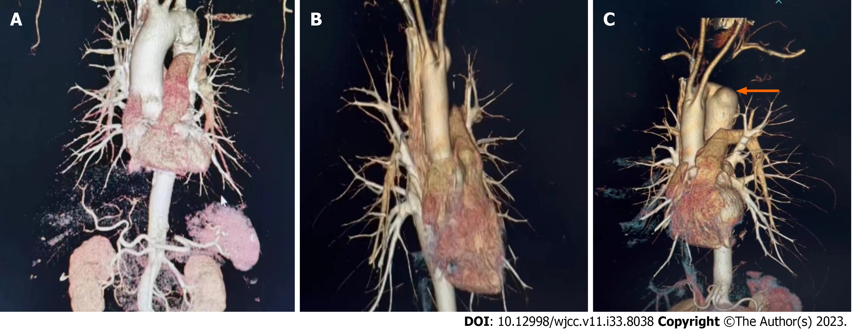
Figure 1 Computed tomographic aortography images.
A: The normal shape of the aorta of this patient; B: The shape of the patient's aorta; C: White arrow showing Kommerell’s diverticulum in the arch of aorta, left subclavian, left cervical spine.
- Citation: Akilu W, Feng Y, Zhang XX, Li SL, Ma XT, Hu M, Cheng C. Carotid-subclavian bypass and endovascular aortic repair of Kommerell’s diverticulum with aberrant left subclavian artery: A case report. World J Clin Cases 2023; 11(33): 8038-8043
- URL: https://www.wjgnet.com/2307-8960/full/v11/i33/8038.htm
- DOI: https://dx.doi.org/10.12998/wjcc.v11.i33.8038


