Copyright
©The Author(s) 2023.
World J Clin Cases. Nov 16, 2023; 11(32): 7888-7894
Published online Nov 16, 2023. doi: 10.12998/wjcc.v11.i32.7888
Published online Nov 16, 2023. doi: 10.12998/wjcc.v11.i32.7888
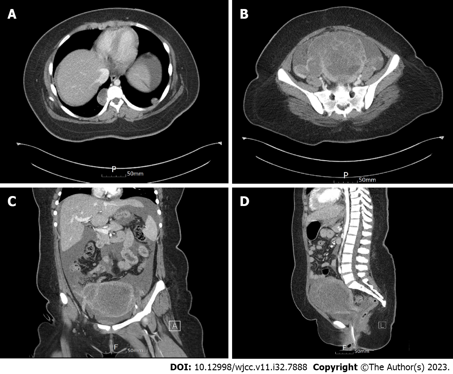
Figure 1 Radiologic findings.
A: Chest computed tomography scan revealed two nodules, suggesting metastatic lesions; B-D: Abdominal computed tomography scan demonstrated a huge mass in the uterine corpus, suggesting a uterine malignancy (B: Axial; C: Coronal; D: Sagittal).
- Citation: Kim NI, Lee JS, Nam JH. Uterine rupture due to adenomyosis in an adolescent: A case report and review of literature. World J Clin Cases 2023; 11(32): 7888-7894
- URL: https://www.wjgnet.com/2307-8960/full/v11/i32/7888.htm
- DOI: https://dx.doi.org/10.12998/wjcc.v11.i32.7888


