Copyright
©The Author(s) 2023.
World J Clin Cases. Nov 6, 2023; 11(31): 7640-7646
Published online Nov 6, 2023. doi: 10.12998/wjcc.v11.i31.7640
Published online Nov 6, 2023. doi: 10.12998/wjcc.v11.i31.7640
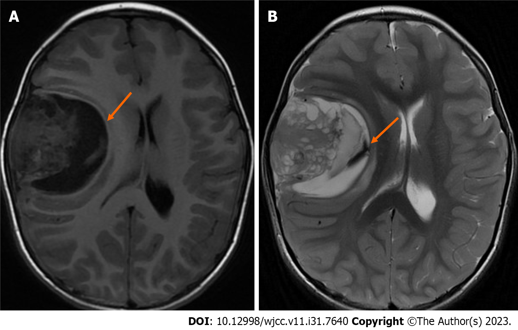
Figure 1 Preoperative nonenhanced magnetic resonance imaging shows a large space-occupying lesion containing cystic and solid components in the right frontotemporal lobe.
A: T1 weighted image; B: T2 weighted image.
- Citation: Huang LJ, Jiao JF, He Q, Luo JW, Guo Y. Ultrafast power Doppler imaging for ischemic encephalopathy: A case report. World J Clin Cases 2023; 11(31): 7640-7646
- URL: https://www.wjgnet.com/2307-8960/full/v11/i31/7640.htm
- DOI: https://dx.doi.org/10.12998/wjcc.v11.i31.7640


