
















©The Author(s) 2023.
World J Clin Cases. Oct 26, 2023; 11(30): 7268-7276
Published online Oct 26, 2023. doi: 10.12998/wjcc.v11.i30.7268
Published online Oct 26, 2023. doi: 10.12998/wjcc.v11.i30.7268
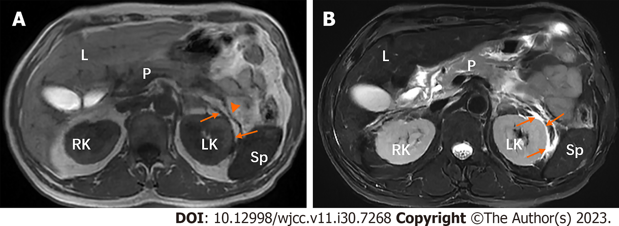
Figure 9 A 52-year-old man with type 2 diabetes mellitus over 10 years with moderately severe acute pancreatitis whose fasting blood glucose level was 15.
48 mmol/L. A and B: In the left perirenal space, there are strips (arrows) of hypointensity on T1-weighted image (A) and hyperintensity on T2-weighted image with fat suppression (B). T1-weighted image (A) also shows the thickened left renal fascia (arrowhead). L: Liver; LK: Left kidney; P: Pancreas; RK: Right kidney; Sp: Spleen.
- Citation: Ni YH, Song LJ, Xiao B. Magnetic resonance imaging for acute pancreatitis in type 2 diabetes patients. World J Clin Cases 2023; 11(30): 7268-7276
- URL: https://www.wjgnet.com/2307-8960/full/v11/i30/7268.htm
- DOI: https://dx.doi.org/10.12998/wjcc.v11.i30.7268