Copyright
©The Author(s) 2023.
World J Clin Cases. Jan 26, 2023; 11(3): 709-718
Published online Jan 26, 2023. doi: 10.12998/wjcc.v11.i3.709
Published online Jan 26, 2023. doi: 10.12998/wjcc.v11.i3.709
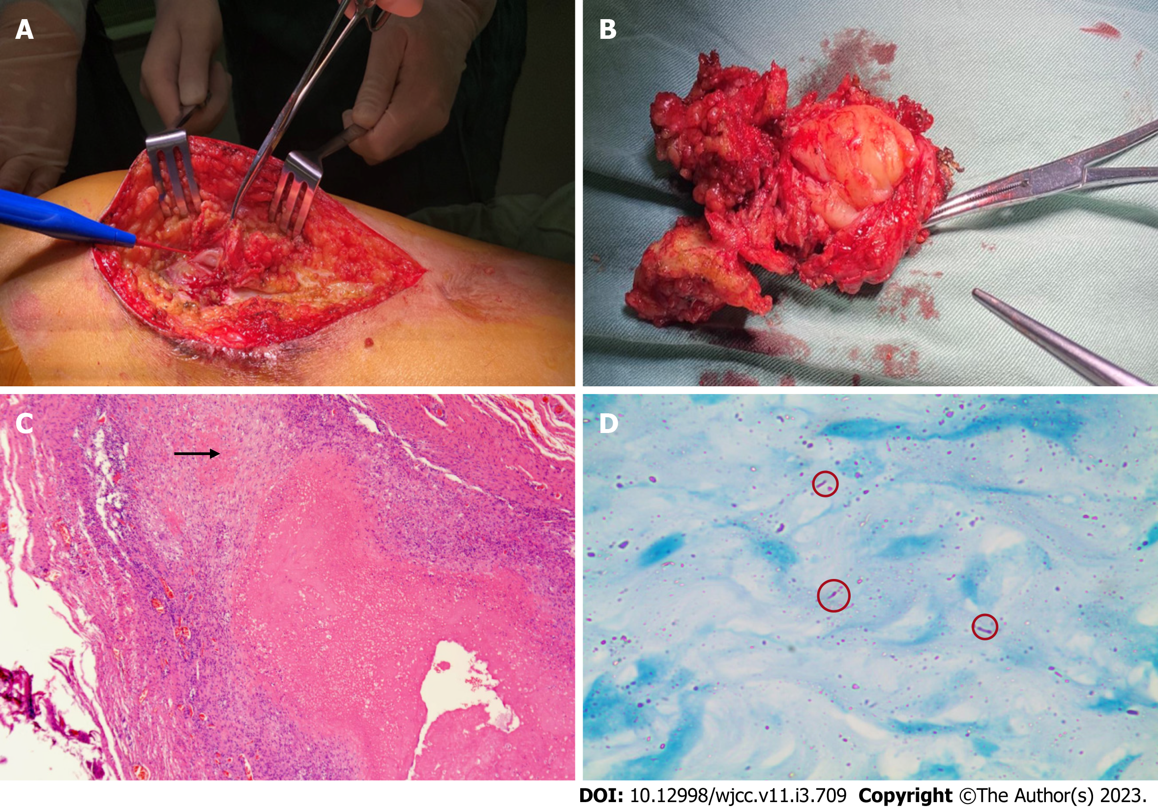
Figure 2 Intraoperative photographs of the left thigh and histopathological examination of resected specimens.
A: Intraoperative photograph; B: Excised specimen (8 cm × 6 cm); C: Granulomas are embedded among the muscle fibers with lymphocyte infiltration and multinucleated giant cell aggregation (× 40); D: The acid-fast bacilli staining test showed scattered, suspicious antacid staining-positive rods (× 1000).
- Citation: He YG, Huang YH, Yi XL, Qian KL, Wang Y, Cheng H, Hu J, Liu Y. Soft tissue tuberculosis detected by next-generation sequencing: A case report and review of literature. World J Clin Cases 2023; 11(3): 709-718
- URL: https://www.wjgnet.com/2307-8960/full/v11/i3/709.htm
- DOI: https://dx.doi.org/10.12998/wjcc.v11.i3.709


