Copyright
©The Author(s) 2023.
World J Clin Cases. Jan 26, 2023; 11(3): 684-691
Published online Jan 26, 2023. doi: 10.12998/wjcc.v11.i3.684
Published online Jan 26, 2023. doi: 10.12998/wjcc.v11.i3.684
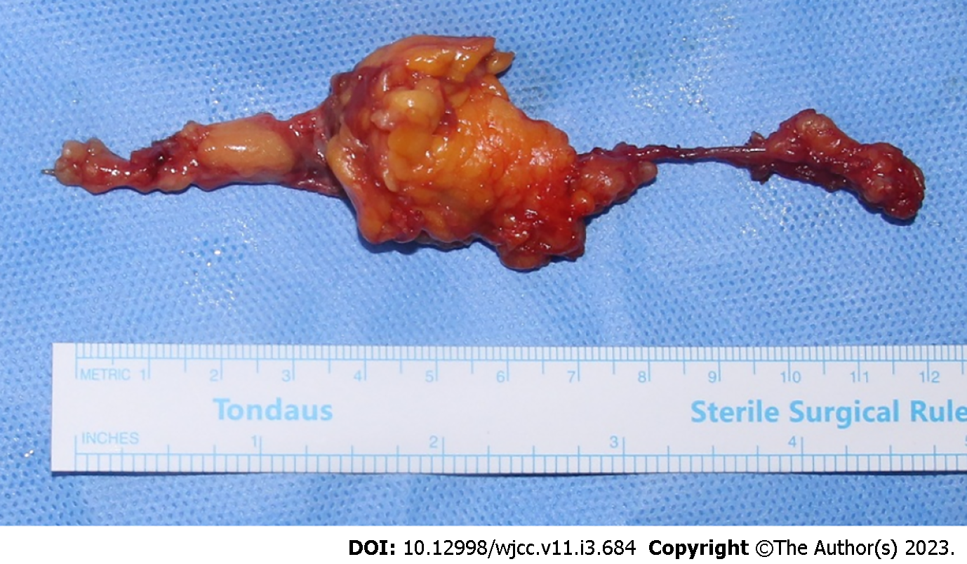
Figure 5 Photograph of the specimen.
A lipomatous mass of 12 cm × 4 cm × 5 cm was excised. It infiltrated the extensor pollicis brevis (EPB) muscle and tendon sheath. Most of the mass burden was located in the muscle portion of the EPB, presenting as yellow tissue with lobulated aspect and muscular fibers throughout.
- Citation: Byeon JY, Hwang YS, Lee JH, Choi HJ. Recurrent intramuscular lipoma at extensor pollicis brevis: A case report. World J Clin Cases 2023; 11(3): 684-691
- URL: https://www.wjgnet.com/2307-8960/full/v11/i3/684.htm
- DOI: https://dx.doi.org/10.12998/wjcc.v11.i3.684


