Copyright
©The Author(s) 2023.
World J Clin Cases. Oct 6, 2023; 11(28): 6920-6930
Published online Oct 6, 2023. doi: 10.12998/wjcc.v11.i28.6920
Published online Oct 6, 2023. doi: 10.12998/wjcc.v11.i28.6920
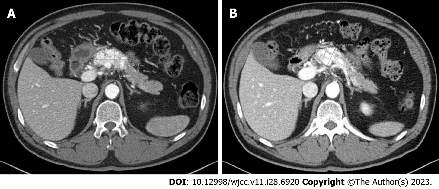
Figure 10 Follow-up abdominal computed tomography scan.
A: On the three-year follow-up computed tomography scan, there was no significant change in the lesion size, which measured 5.7 cm × 3.3 cm; B: On the nine-year follow-up scale, the lesion size remained at 6.0 cm × 3.0 cm.
- Citation: Shin SH, Cho CK, Yu SY. Pancreatic arteriovenous malformation treated with transcatheter arterial embolization: Two case reports and review of literature. World J Clin Cases 2023; 11(28): 6920-6930
- URL: https://www.wjgnet.com/2307-8960/full/v11/i28/6920.htm
- DOI: https://dx.doi.org/10.12998/wjcc.v11.i28.6920


