Copyright
©The Author(s) 2023.
World J Clin Cases. Oct 6, 2023; 11(28): 6920-6930
Published online Oct 6, 2023. doi: 10.12998/wjcc.v11.i28.6920
Published online Oct 6, 2023. doi: 10.12998/wjcc.v11.i28.6920
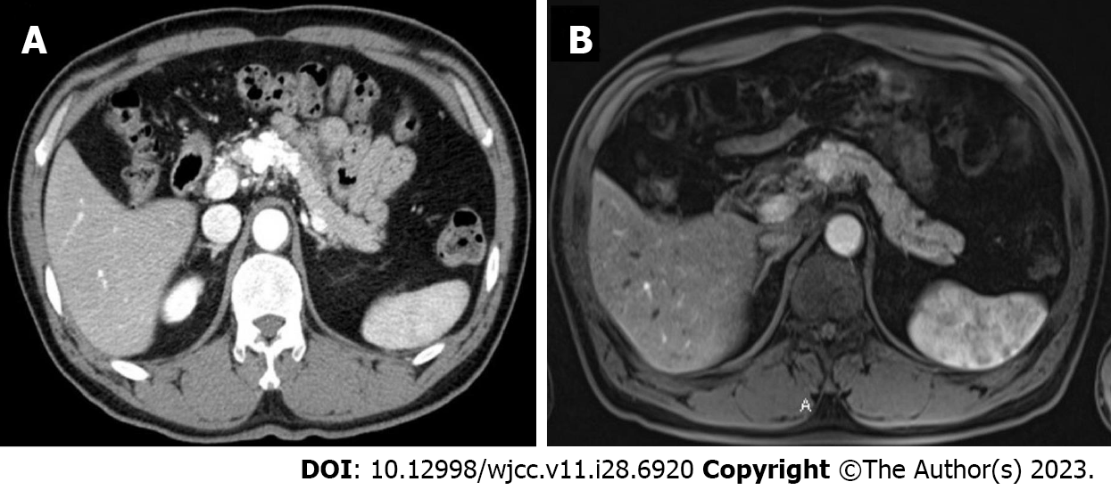
Figure 2 Abdominal computed tomography and magnetic resonance imaging before transcatheter arterial embolization (case 2).
A: Computed tomography image showing a 5.2 cm × 4.0 cm enhancing tortuous, tubular hypervascular lesion in the pancreas neck, and body; B: Magnetic resonance imaging image showing a 2 cm multilobulated and irregular hypervascular lesion in the pancreatic neck and body with peripancreatic infiltration.
- Citation: Shin SH, Cho CK, Yu SY. Pancreatic arteriovenous malformation treated with transcatheter arterial embolization: Two case reports and review of literature. World J Clin Cases 2023; 11(28): 6920-6930
- URL: https://www.wjgnet.com/2307-8960/full/v11/i28/6920.htm
- DOI: https://dx.doi.org/10.12998/wjcc.v11.i28.6920


