Copyright
©The Author(s) 2023.
World J Clin Cases. Oct 6, 2023; 11(28): 6864-6870
Published online Oct 6, 2023. doi: 10.12998/wjcc.v11.i28.6864
Published online Oct 6, 2023. doi: 10.12998/wjcc.v11.i28.6864
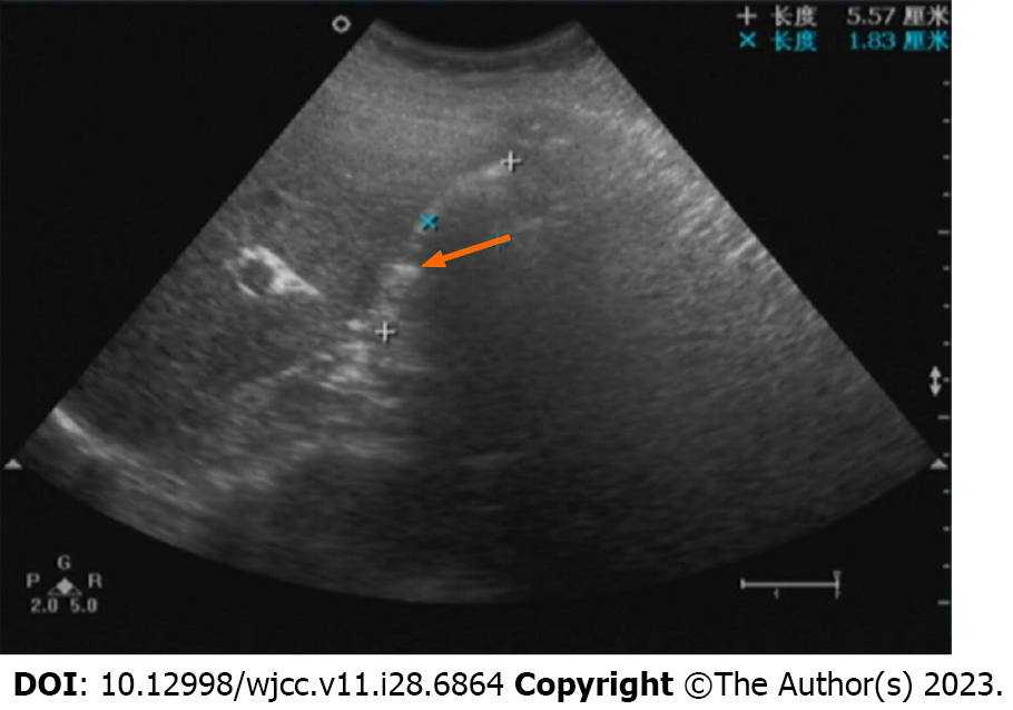
Figure 1 Ultrasound diagnosis before surgery.
Transverse view of the liver showing the gallbladder was filled with a strong hyperechoic mass of uncertain size (arrow).
- Citation: Sun HJ, Ge F, Si Y, Wang Z, Sun HB. Importance of accurate diagnosis of congenital agenesis of the gallbladder from atypical gallbladder stone presentations: A case report. World J Clin Cases 2023; 11(28): 6864-6870
- URL: https://www.wjgnet.com/2307-8960/full/v11/i28/6864.htm
- DOI: https://dx.doi.org/10.12998/wjcc.v11.i28.6864


