Copyright
©The Author(s) 2023.
World J Clin Cases. Sep 26, 2023; 11(27): 6653-6663
Published online Sep 26, 2023. doi: 10.12998/wjcc.v11.i27.6653
Published online Sep 26, 2023. doi: 10.12998/wjcc.v11.i27.6653
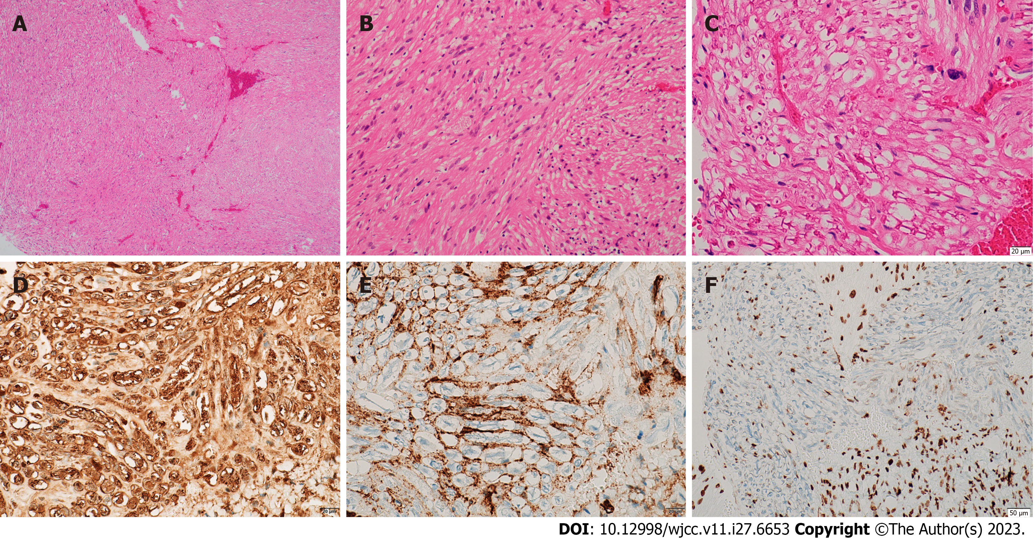
Figure 2 Microscopic features of neurofibroma.
A-C: Histopathological examination with toluidine blue and hematoxylin and eosin staining showing spindle fiber and fibroblast-like cell proliferation with slight nuclear enlargement. The nuclei were short and spindle-shaped, the cytoplasm was richly stained red, and axons were locally visible; D: Immunohistochemistry positive for S-100 protein; E: Immunohistochemistry positive for CD34; F: Immunohistochemistry positive for H3K27ME3.
- Citation: Zhang Z, Hong X, Wang F, Ye X, Yao YD, Yin Y, Yang HY. Solitary intraosseous neurofibroma in the mandible mimicking a cystic lesion: A case report and review of literature. World J Clin Cases 2023; 11(27): 6653-6663
- URL: https://www.wjgnet.com/2307-8960/full/v11/i27/6653.htm
- DOI: https://dx.doi.org/10.12998/wjcc.v11.i27.6653


