Copyright
©The Author(s) 2023.
World J Clin Cases. Sep 26, 2023; 11(27): 6579-6586
Published online Sep 26, 2023. doi: 10.12998/wjcc.v11.i27.6579
Published online Sep 26, 2023. doi: 10.12998/wjcc.v11.i27.6579
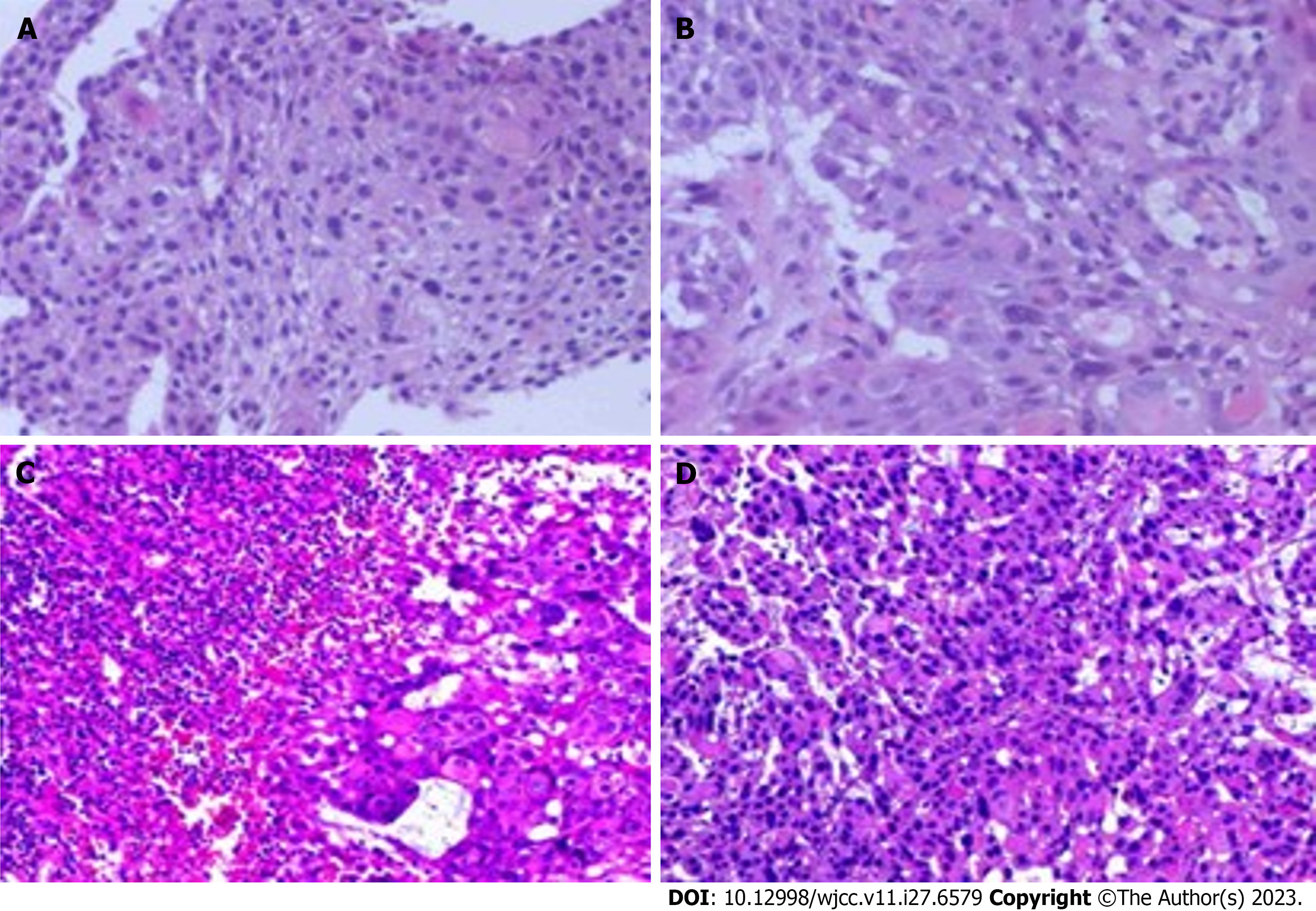
Figure 2 Histopathological examination by hematoxylin-eosin staining.
A and B: Heterogeneous proliferation of tissue squamous epithelium. The heterogeneous cells break through the basement membrane and infiltrate below the mesenchyme (40 ×, 200 ×); C and D: The squamous epithelial cells were markedly heterogeneous and showed infiltrative growth, and a little lymphoid tissue was seen in their periphery (40 ×, 200 ×).
- Citation: Chen SC, Ma DH, Zhong JJ. Combination therapy with toripalimab and anlotinib in advanced esophageal squamous cell carcinoma: A case report. World J Clin Cases 2023; 11(27): 6579-6586
- URL: https://www.wjgnet.com/2307-8960/full/v11/i27/6579.htm
- DOI: https://dx.doi.org/10.12998/wjcc.v11.i27.6579


