Copyright
©The Author(s) 2023.
World J Clin Cases. Sep 26, 2023; 11(27): 6573-6578
Published online Sep 26, 2023. doi: 10.12998/wjcc.v11.i27.6573
Published online Sep 26, 2023. doi: 10.12998/wjcc.v11.i27.6573
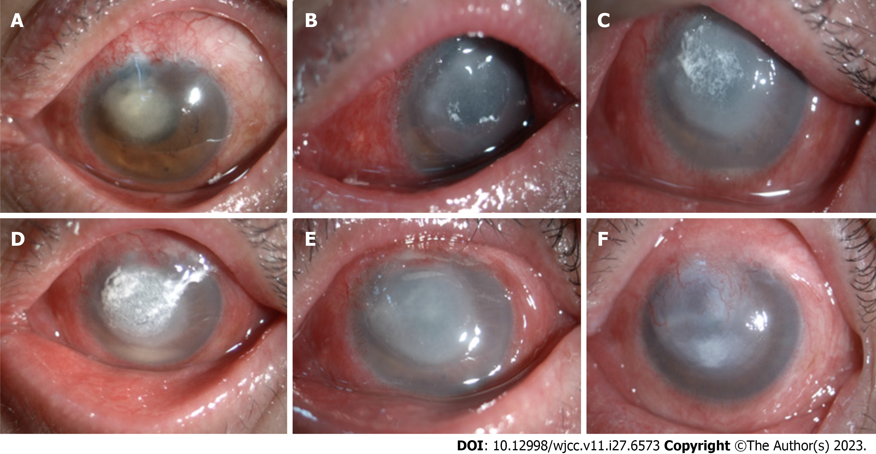
Figure 2 The course of the fungal corneal infection.
A: The 4th wk after the operation. There is a grey infiltrate with irregular white edges on the cornea; B: The 5th wk after operation. A corneal ulcer has formed; C: The 7th wk after operation. A hypopyon appears to have occurred; D: The 9th wk after operation. The corneal ulcer and hypopyon is still present; E: The 11th wk after operation. The corneal ulcer appears to be improving, the hypopyon has absorbed; F: The 16th wk after operation. The corneal ulcer is repaired, a leucoma appears to have formed.
- Citation: Zhao J, Xu HT, Yin Y, Li YX, Zheng YJ. Fungal corneal ulcer after repair of an overhanging filtering bleb: A case report. World J Clin Cases 2023; 11(27): 6573-6578
- URL: https://www.wjgnet.com/2307-8960/full/v11/i27/6573.htm
- DOI: https://dx.doi.org/10.12998/wjcc.v11.i27.6573


