Copyright
©The Author(s) 2023.
World J Clin Cases. Sep 26, 2023; 11(27): 6573-6578
Published online Sep 26, 2023. doi: 10.12998/wjcc.v11.i27.6573
Published online Sep 26, 2023. doi: 10.12998/wjcc.v11.i27.6573
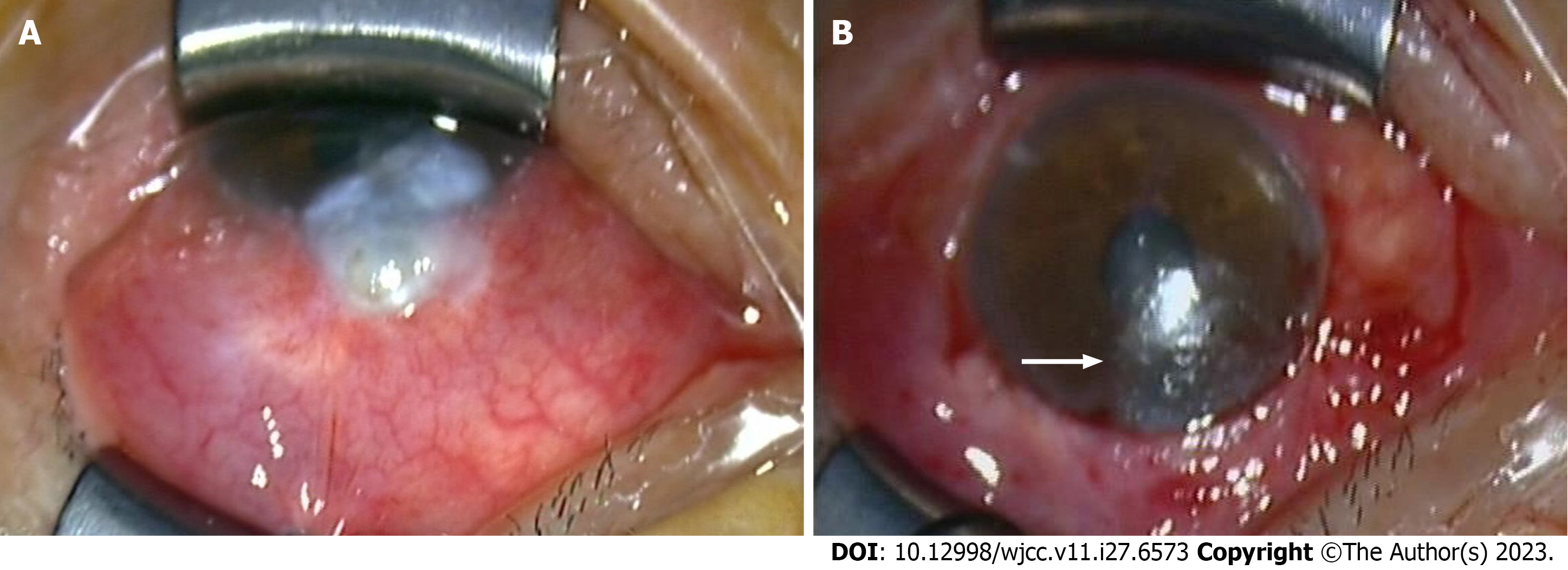
Figure 1 Preoperative and postoperative photograph of the eye.
A: Preoperative photograph: A prolapsed filtering bleb invading the transparent cornea and approaching the pupil area is shown; B: Postoperative photograph taken by operating microscope: The repaired filtering bleb is shown by the white arrow in the lower part of the photo; a corneal epithelial defect remained in the prolapsed area of the filtering bleb.
- Citation: Zhao J, Xu HT, Yin Y, Li YX, Zheng YJ. Fungal corneal ulcer after repair of an overhanging filtering bleb: A case report. World J Clin Cases 2023; 11(27): 6573-6578
- URL: https://www.wjgnet.com/2307-8960/full/v11/i27/6573.htm
- DOI: https://dx.doi.org/10.12998/wjcc.v11.i27.6573


