Copyright
©The Author(s) 2023.
World J Clin Cases. Sep 26, 2023; 11(27): 6558-6564
Published online Sep 26, 2023. doi: 10.12998/wjcc.v11.i27.6558
Published online Sep 26, 2023. doi: 10.12998/wjcc.v11.i27.6558
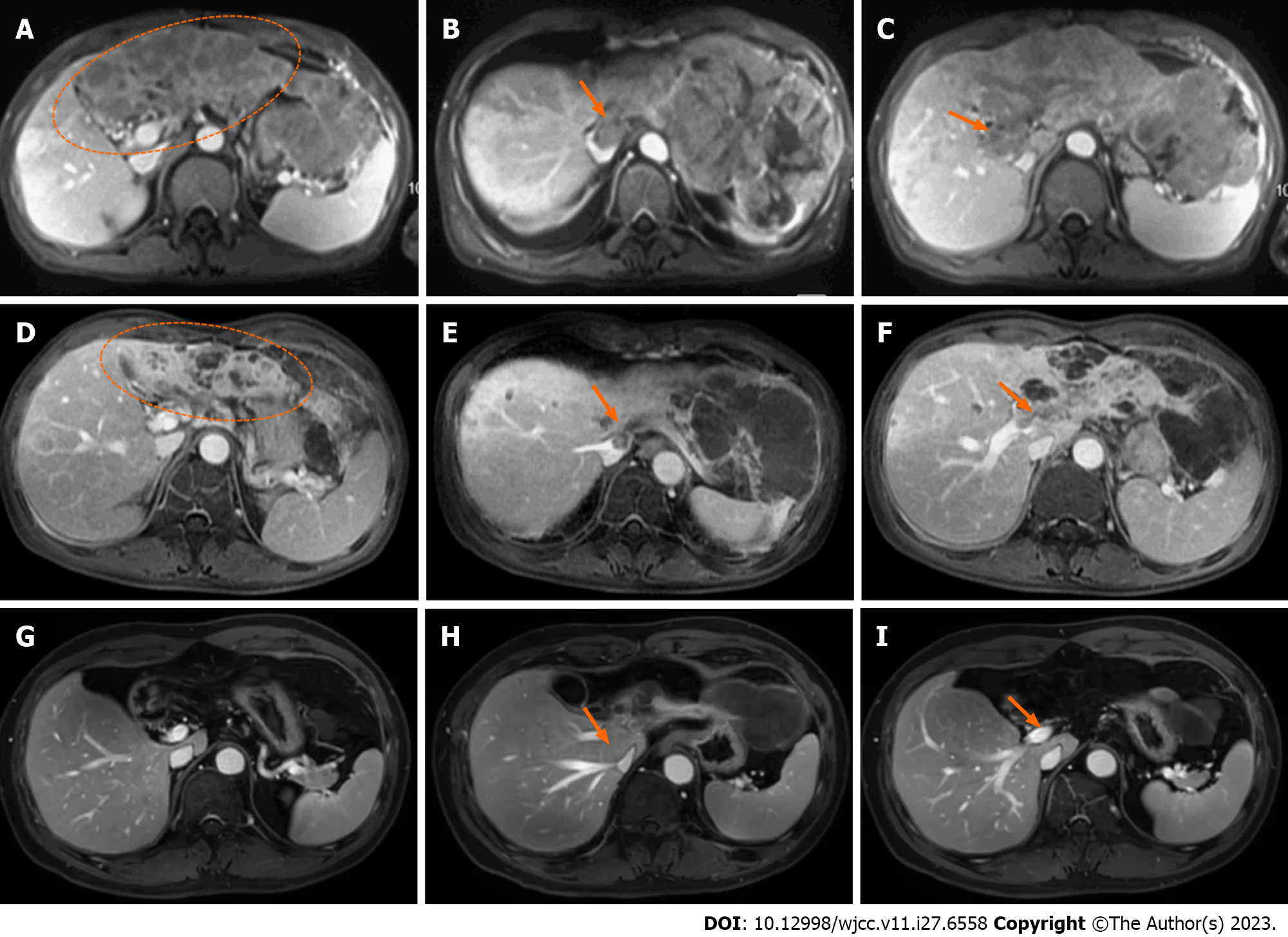
Figure 1 The dynamic contrast-enhanced magnetic resonance imaging showed hepatocellular carcinoma, inferior vena cava tumor thrombus and main portal vein tumor thrombus before and after therapy.
A-C: Images before treatment (March 2018), the liver tumor (A, ellipse): Inferior vena cava tumor thrombus (IVCTT) (B, arrow), the main portal vein tumor thrombus (MPVTT) (C, arrow); D-F: Images of 2 mo after radiotherapy (June 2018), partial remission of the tumor (D, ellipse), partial remission of the IVCTT (E, arrow), and partial remission of the MPVTT (F, arrow); G-I: Images of 36 mo after radiotherapy (May 2021), complete remission of tumor (G), complete remission of IVCTT (H), and complete remission of MPVTT (I).
- Citation: Zhao Y, He GS, Li G. Triplet regimen as a novel modality for advanced unresectable hepatocellular carcinoma: A case report and review of literature. World J Clin Cases 2023; 11(27): 6558-6564
- URL: https://www.wjgnet.com/2307-8960/full/v11/i27/6558.htm
- DOI: https://dx.doi.org/10.12998/wjcc.v11.i27.6558


