Copyright
©The Author(s) 2023.
World J Clin Cases. Sep 26, 2023; 11(27): 6551-6557
Published online Sep 26, 2023. doi: 10.12998/wjcc.v11.i27.6551
Published online Sep 26, 2023. doi: 10.12998/wjcc.v11.i27.6551
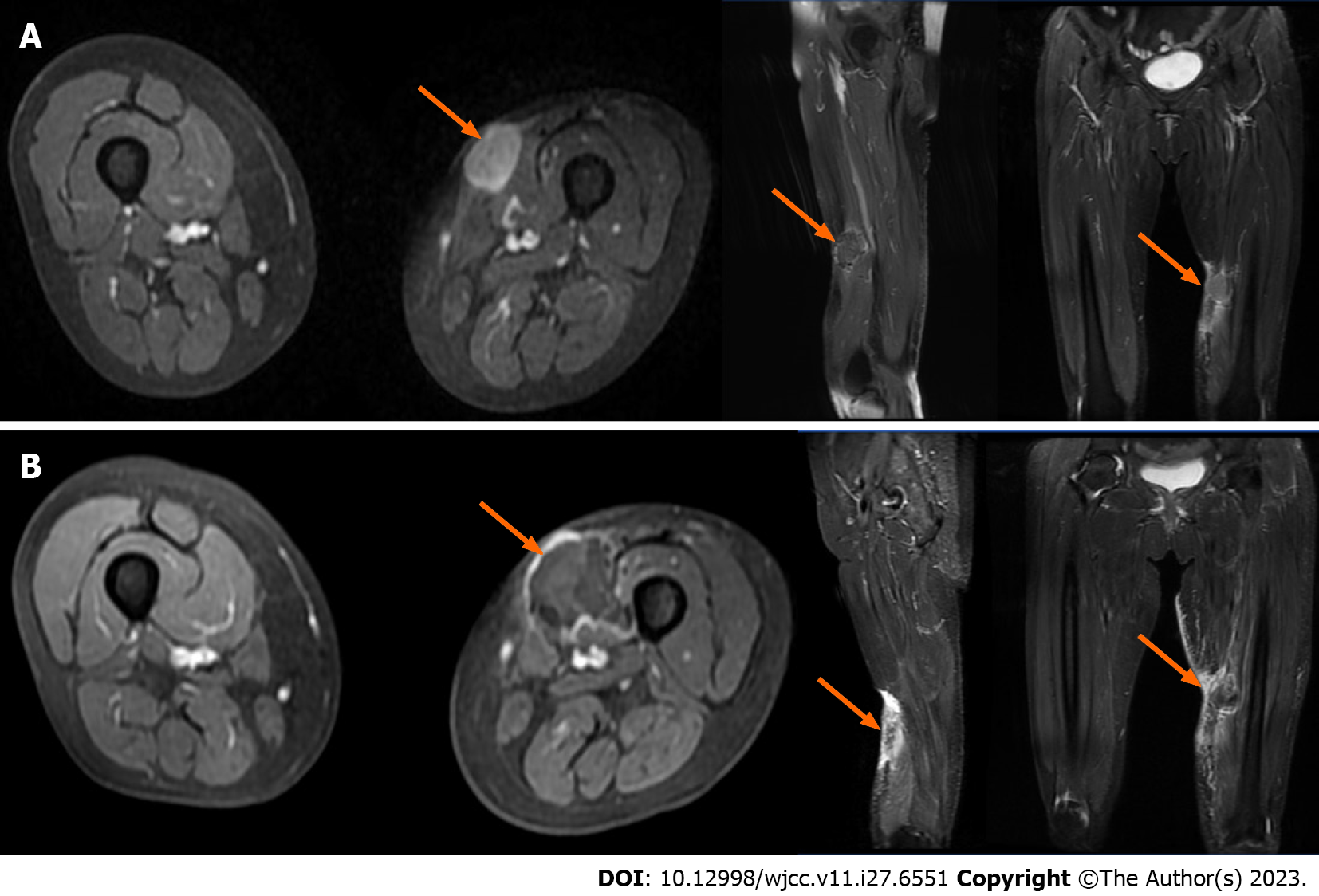
Figure 4 Comparison of magnetic resonance imaging of lower limbs before and after high-intensity focused ultrasound treatment.
A and B: Enhanced magnetic resonance imaging (MRI) of the left thigh. A: Before high-intensity focused ultrasound (HIFU) (March 1, 2021): On the coronal, sagittal and cross-sectional MRI of the left thigh, an oval signal nodule with slightly longer T1 and slightly longer T2 (about 31 mm × 26 mm × 30 mm) was found under the skin of the anterior medial side of the middle part of the left thigh (arrows). After enhancement, it was obviously slightly uneven, and the surrounding soft tissues were edema; B: After HIFU (March 12, 2021): On the coronal, sagittal and cross-sectional MRI of the left thigh, the oval signal nodule with slightly longer T1 and slightly longer T2 (about 28 mm × 30 mm × 25 mm) is located under the skin of the anterior medial side of the middle part of the left thigh, and its boundary with subcutaneous fat and adjacent muscle tissue is unclear (arrows).
- Citation: Zhu YQ, Zhao GC, Zheng CX, Yuan L, Yuan GB. Managing spindle cell sarcoma with surgery and high-intensity focused ultrasound: A case report. World J Clin Cases 2023; 11(27): 6551-6557
- URL: https://www.wjgnet.com/2307-8960/full/v11/i27/6551.htm
- DOI: https://dx.doi.org/10.12998/wjcc.v11.i27.6551


