Copyright
©The Author(s) 2023.
World J Clin Cases. Sep 26, 2023; 11(27): 6543-6550
Published online Sep 26, 2023. doi: 10.12998/wjcc.v11.i27.6543
Published online Sep 26, 2023. doi: 10.12998/wjcc.v11.i27.6543
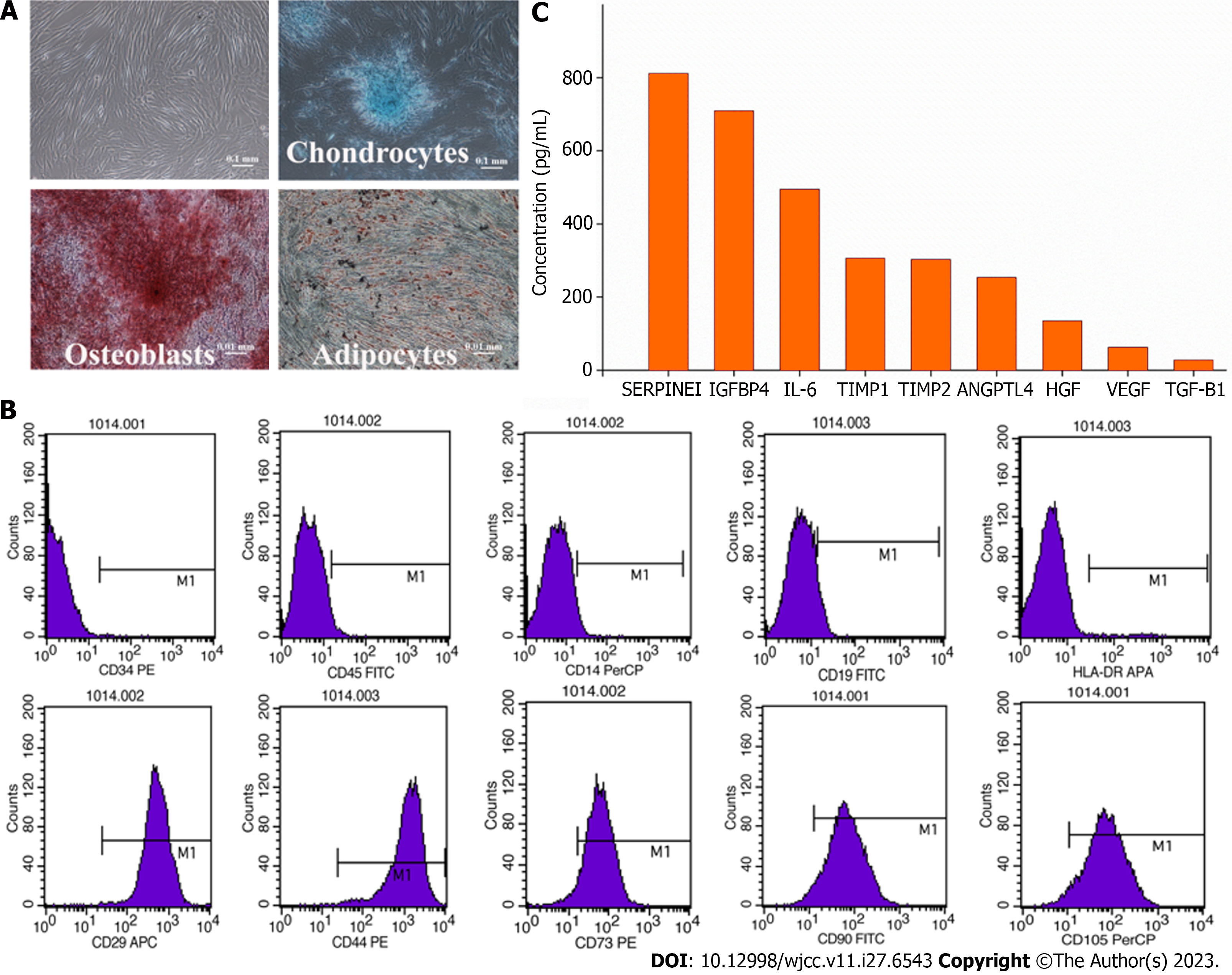
Figure 2 Quality control of amniotic membrane mesenchymal stromal cells and the mesenchymal stromal cell-secretome.
A: Cell characterization and differentiation potential. Amniotic membrane-mesenchymal stromal cells were characterized by their fibroblast-like morphology and their differentiation potential towards osteoblasts, chondrocytes, and adipocytes in special induction medium; B: Surface markers. The mniotic membrane-mesenchymal stromal cells showed a high expression of CD29, CD44, CD73, CD90, CD105 and low expression of CD14, CD19, CD34, CD45, and HLA-DR markers; C: Cytokine concentration. ANGPTL4: Angiopoietin-like 4; HGF: Hepatocyte growth factor; IGFBP4: Insulin-like growth factor binding protein 4; IL-6: Interleukin-6; TIMP1: Tissue inhibitors of metalloproteinase 1; SERPINE1: Serine protease inhibitor clade E member 1; TGF-β1: Transforming growth factor-beta 1; TIMP-2: Tissue inhibitors of metalloproteinase 2; VEGF: Vascular endothelial growth factor.
- Citation: Lin FH, Yang YX, Wang YJ, Subbiah SK, Wu XY. Amniotic membrane mesenchymal stromal cell-derived secretome in the treatment of acute ischemic stroke: A case report. World J Clin Cases 2023; 11(27): 6543-6550
- URL: https://www.wjgnet.com/2307-8960/full/v11/i27/6543.htm
- DOI: https://dx.doi.org/10.12998/wjcc.v11.i27.6543


