Copyright
©The Author(s) 2023.
World J Clin Cases. Sep 26, 2023; 11(27): 6363-6373
Published online Sep 26, 2023. doi: 10.12998/wjcc.v11.i27.6363
Published online Sep 26, 2023. doi: 10.12998/wjcc.v11.i27.6363
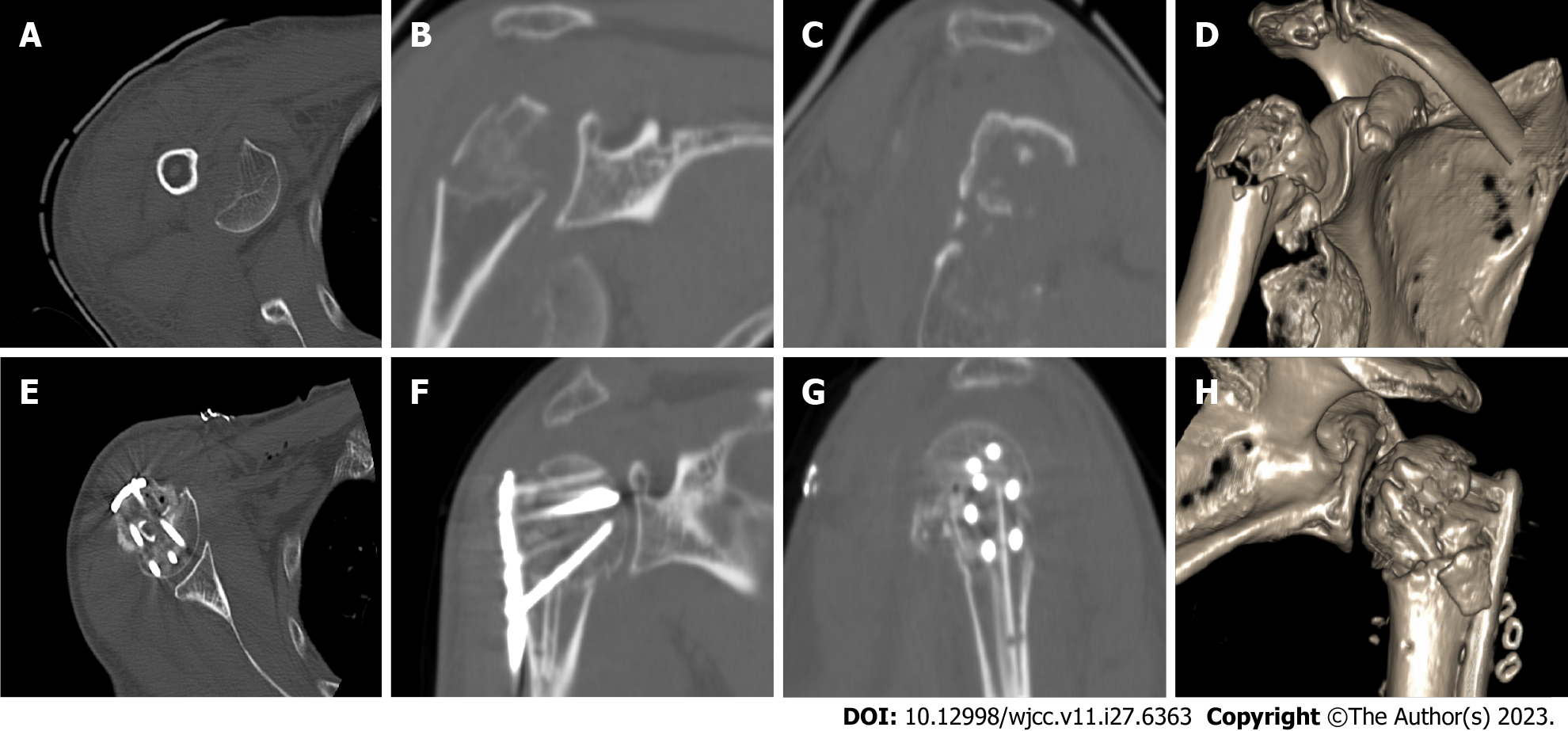
Figure 3 Computed tomography images of a typical case.
A: Preoperative computed tomography (CT) image of the right proximal humerus in the horizontal position; B: Preoperative CT image of the right proximal humerus in the coronal position; C: Preoperative CT image of the right proximal humerus in the sagittal position; D: Three-dimensional CT image before reconstruction of the right proximal humerus; E: Postoperative CT image of the right proximal humerus in the horizontal position; F: Postoperative CT image of the right proximal humerus in the coronal position; G: Postoperative CT image of the right proximal humerus in the sagittal position; H: Three-dimensional CT image after reconstruction of the right proximal humerus.
- Citation: Liu N, Wang BG, Zhang LF. Treatment of proximal humeral fractures accompanied by medial calcar fractures using fibular autografts: A retrospective, comparative cohort study. World J Clin Cases 2023; 11(27): 6363-6373
- URL: https://www.wjgnet.com/2307-8960/full/v11/i27/6363.htm
- DOI: https://dx.doi.org/10.12998/wjcc.v11.i27.6363


