Copyright
©The Author(s) 2023.
World J Clin Cases. Sep 16, 2023; 11(26): 6289-6297
Published online Sep 16, 2023. doi: 10.12998/wjcc.v11.i26.6289
Published online Sep 16, 2023. doi: 10.12998/wjcc.v11.i26.6289
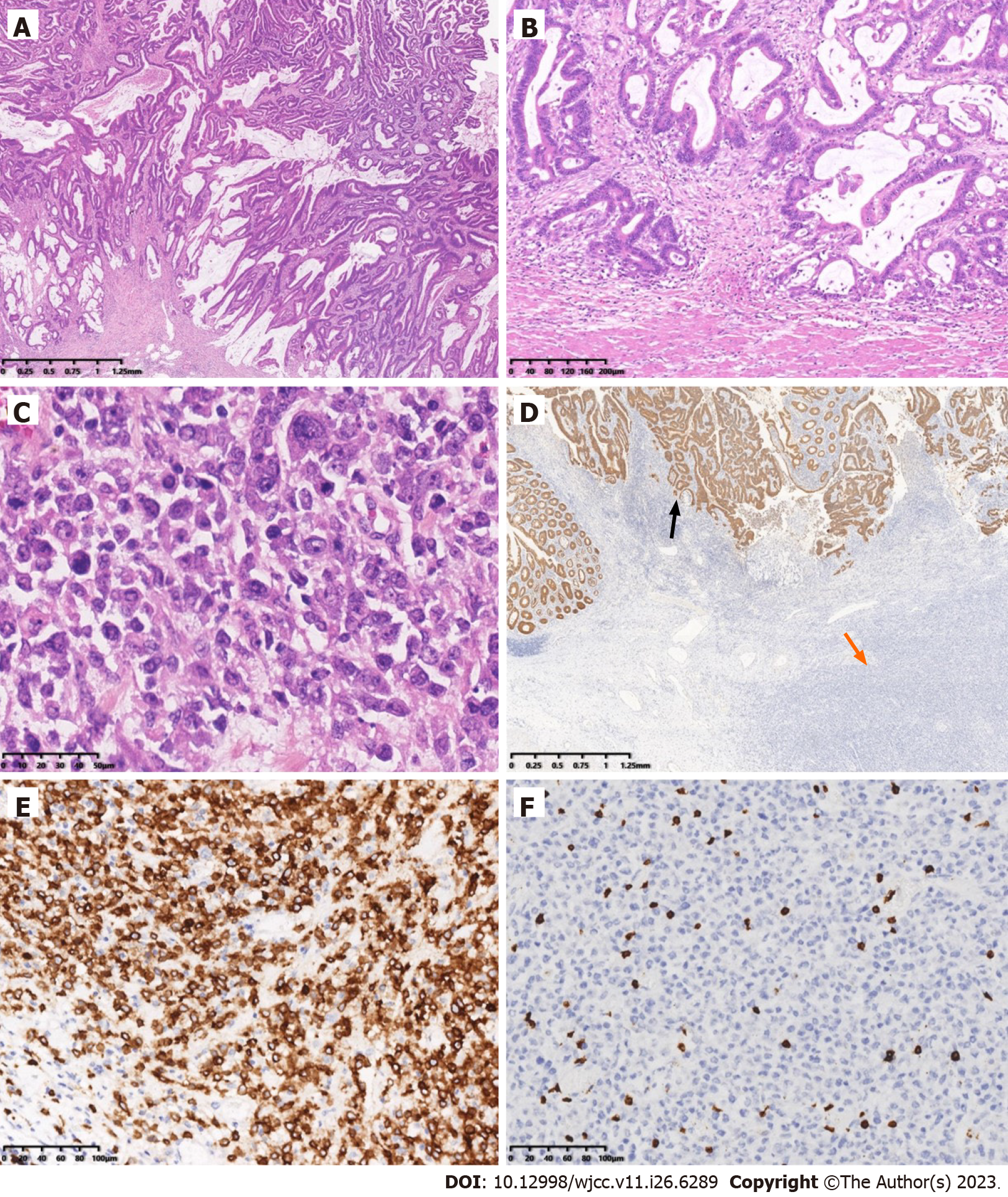
Figure 3 Microscopic examination and immunohistochemistry.
A: Histopathology of one component of the mass was a moderately differentiated adenocarcinoma having a mucinous component arising from a tubulovillous adenoma (top) (Hematoxylin and eosin (HE), original magnification 20×); B: Adenocarcinoma invaded the muscularis propria (HE, original magnification 100×); C: The other component was diffusely medium-to-large lymphocytes, with uniform morphology, frequent mitosis and obvious cell atypia (HE, original magnification 400×); D: Immunohistochemistry of adenocarcinoma was strongly positive for cytokeratin (black arrow), while the atypical lymphocytes were negative (orange arrow) (EnVision method, original magnification 20×); E: Immunohistochemistry of the atypical lymphocytes was strongly and diffusely positive for CD20 (EnVision method, original magnification 200×); F: Immunohistochemistry of the atypical lymphocytes was negative for CD3 (EnVision method, original magnification 200×).
- Citation: Jiang M, Yuan XP. Collision tumor of primary malignant lymphoma and adenocarcinoma in the colon diagnosed by molecular pathology: A case report and literature review. World J Clin Cases 2023; 11(26): 6289-6297
- URL: https://www.wjgnet.com/2307-8960/full/v11/i26/6289.htm
- DOI: https://dx.doi.org/10.12998/wjcc.v11.i26.6289


