Copyright
©The Author(s) 2023.
World J Clin Cases. Sep 16, 2023; 11(26): 6289-6297
Published online Sep 16, 2023. doi: 10.12998/wjcc.v11.i26.6289
Published online Sep 16, 2023. doi: 10.12998/wjcc.v11.i26.6289
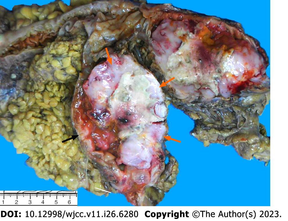
Figure 1 Macroscopic examination.
The resected specimen presented as a circumferential ulcerative mass on the cecal mucosa adjacent to the ileocecal valve. The mass appeared to be comprised of two tumors. The upper-right portion of the mass had a crater-like appearance with a necrotic-appearing central area (orange arrow), whereas the remaining part of the mass had a polypoid, hard and grayish-white aspect (black arrow).
- Citation: Jiang M, Yuan XP. Collision tumor of primary malignant lymphoma and adenocarcinoma in the colon diagnosed by molecular pathology: A case report and literature review. World J Clin Cases 2023; 11(26): 6289-6297
- URL: https://www.wjgnet.com/2307-8960/full/v11/i26/6289.htm
- DOI: https://dx.doi.org/10.12998/wjcc.v11.i26.6289


