Copyright
©The Author(s) 2023.
World J Clin Cases. Sep 16, 2023; 11(26): 6194-6199
Published online Sep 16, 2023. doi: 10.12998/wjcc.v11.i26.6194
Published online Sep 16, 2023. doi: 10.12998/wjcc.v11.i26.6194
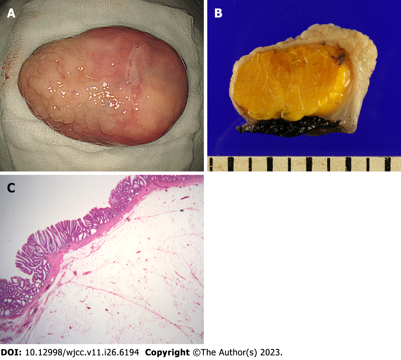
Figure 3 Pathologic findings.
A: Macroscopically, a yellow lipoma was observed in the submucosa and a laterally spreading tumor was observed on the mucosal surface; B: The black color at the bottom of the lipoma was not a burnt area due to electrical thermal injury, but a stain indicated the border of the bottom for pathological evaluation; C: Microscopically, the mucosal lesion on the surface of the lipoma showed tubulovillous adenoma with low-grade dysplasia (H&E staining, magnification × 12.5).
- Citation: Bae JY, Kim HK, Kim YJ, Kim SW, Lee Y, Ryu CB, Lee MS. Large colonic lipoma with a laterally spreading tumor treated by endoscopic submucosal dissection: A case report. World J Clin Cases 2023; 11(26): 6194-6199
- URL: https://www.wjgnet.com/2307-8960/full/v11/i26/6194.htm
- DOI: https://dx.doi.org/10.12998/wjcc.v11.i26.6194


