Copyright
©The Author(s) 2023.
World J Clin Cases. Aug 26, 2023; 11(24): 5762-5771
Published online Aug 26, 2023. doi: 10.12998/wjcc.v11.i24.5762
Published online Aug 26, 2023. doi: 10.12998/wjcc.v11.i24.5762
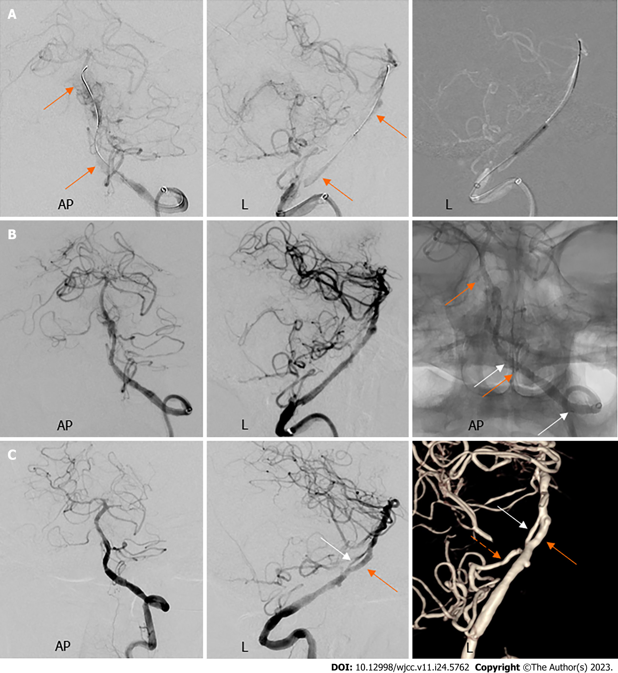
Figure 6 Cerebral angiography of left vertebral artery before balloon dilatation, after stent implantation and at three months postoperatively.
A: Digital subtraction angiography (DSA) before balloon dilatation showed the subintimal canal (orange arrows); B: Angiography revealed reconstruction of the left vertebral artery after implantation of two stents, with four stent markers visible on fluoroscopy (orange and white arrows); C: Follow-up DSA revealed that a residual true lumen (white arrows) was seen in parallel with the subintimal canal (orange arrows) far from the left anterior inferior cerebellar artery (orange dashed arrows), giving the impression of fenestration. AP: Anterior-posterior; L: Lateral.
- Citation: Fu JF, Zhang XL, Lee SY, Zhang FM, You JS. Subintimal recanalization for non-acute occlusion of intracranial vertebral artery in an emergency endovascular procedure: A case report. World J Clin Cases 2023; 11(24): 5762-5771
- URL: https://www.wjgnet.com/2307-8960/full/v11/i24/5762.htm
- DOI: https://dx.doi.org/10.12998/wjcc.v11.i24.5762


