Copyright
©The Author(s) 2023.
World J Clin Cases. Aug 26, 2023; 11(24): 5762-5771
Published online Aug 26, 2023. doi: 10.12998/wjcc.v11.i24.5762
Published online Aug 26, 2023. doi: 10.12998/wjcc.v11.i24.5762
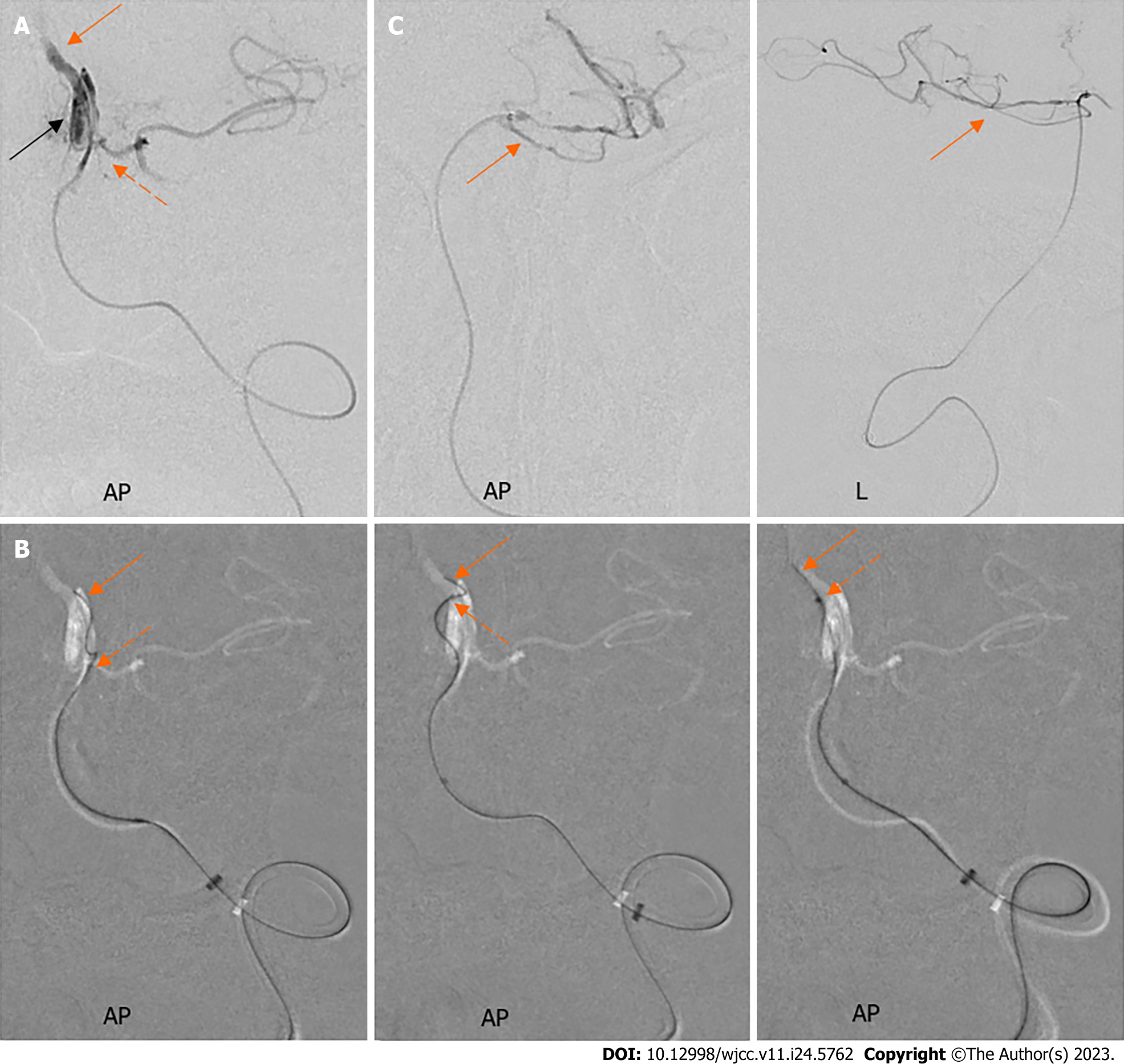
Figure 5 The subintimal microwire re-entered the true lumen.
A: The microcatheter angiography at the level of anterior inferior cerebellar artery (AICA) orifice revealed the true lumen’s position (orange arrow), the AICA branch (the orange dashed arrow), and the subintimal “tubular” dissection (black arrow); B: Under the guidance of a roadmap, the microwire (orange arrows) and the microcatheter (the orange dashed arrows) were manipulated to re-enter the true lumen; C: The microcatheter angiography showed the microwire was advanced to the superior cerebellar artery (orange arrows). AP: anterior-posterior; L: lateral.
- Citation: Fu JF, Zhang XL, Lee SY, Zhang FM, You JS. Subintimal recanalization for non-acute occlusion of intracranial vertebral artery in an emergency endovascular procedure: A case report. World J Clin Cases 2023; 11(24): 5762-5771
- URL: https://www.wjgnet.com/2307-8960/full/v11/i24/5762.htm
- DOI: https://dx.doi.org/10.12998/wjcc.v11.i24.5762


