Copyright
©The Author(s) 2023.
World J Clin Cases. Aug 26, 2023; 11(24): 5762-5771
Published online Aug 26, 2023. doi: 10.12998/wjcc.v11.i24.5762
Published online Aug 26, 2023. doi: 10.12998/wjcc.v11.i24.5762
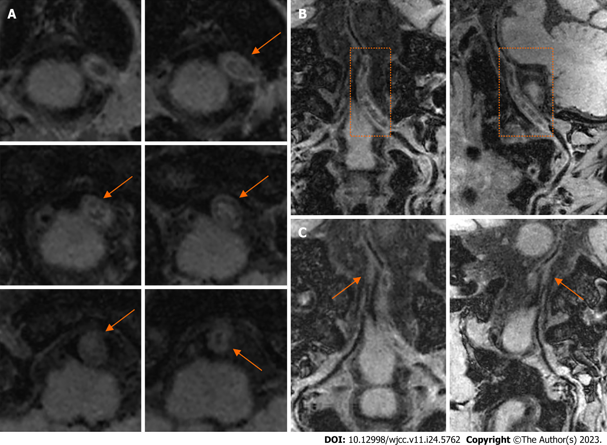
Figure 3 The high-resolution magnetic resonance imaging of bilateral vertebral arteries.
A: The T1-weighted sequence of several axial levels of the left intracranial vertebral artery (VA) showed mixed equal, slightly low, and slightly high signal intensity in the occluded VA lumen, with obvious wall thickening (orange arrows); B: The curved planar reformation (CPR) images of left VA showed long-range thrombosis, lumen occlusion, and wall thickening (orange dashed zones); C: The CPR images showed occlusion of the right intracranial VA (orange arrows).
- Citation: Fu JF, Zhang XL, Lee SY, Zhang FM, You JS. Subintimal recanalization for non-acute occlusion of intracranial vertebral artery in an emergency endovascular procedure: A case report. World J Clin Cases 2023; 11(24): 5762-5771
- URL: https://www.wjgnet.com/2307-8960/full/v11/i24/5762.htm
- DOI: https://dx.doi.org/10.12998/wjcc.v11.i24.5762


