Copyright
©The Author(s) 2023.
World J Clin Cases. Aug 16, 2023; 11(23): 5538-5546
Published online Aug 16, 2023. doi: 10.12998/wjcc.v11.i23.5538
Published online Aug 16, 2023. doi: 10.12998/wjcc.v11.i23.5538
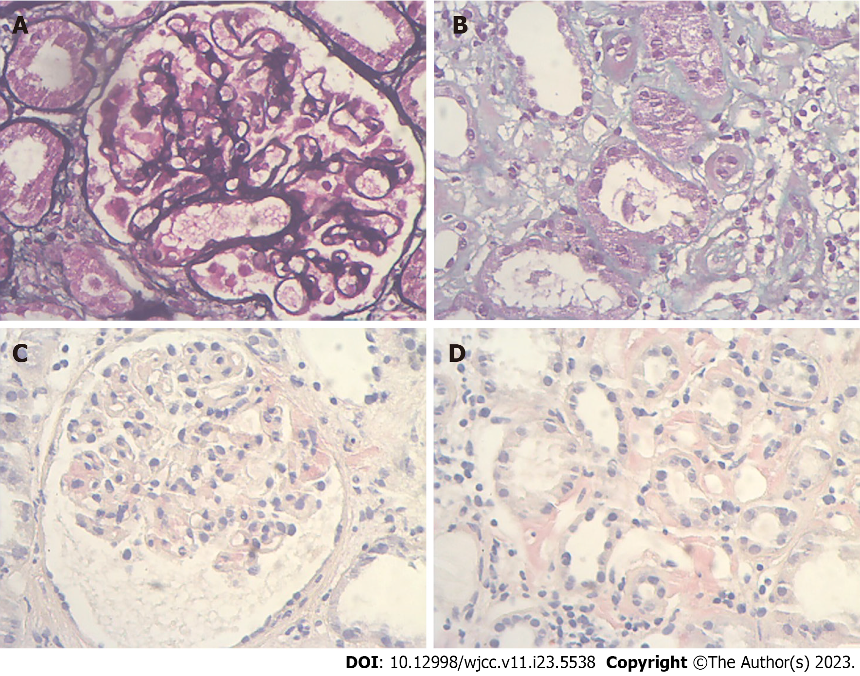
Figure 1 Light microscope images of kidney biopsy.
A: Thickened glomerular basement membrane (PASM stain, 400×); B: Interstitial renal fibrosis (Masson, 400×); C: Positivetive Congo red staining in the glomerular mesangial area (Congo red stain, 400×); D: Positivetive Congo red staining in the renal interstitium (Congo red stain, 400×).
- Citation: Zhang J, Wang X, Zou GM, Li JY, Li WG. Membranous nephropathy with systemic light-chain amyloidosis of remission after rituximab therapy: A case report. World J Clin Cases 2023; 11(23): 5538-5546
- URL: https://www.wjgnet.com/2307-8960/full/v11/i23/5538.htm
- DOI: https://dx.doi.org/10.12998/wjcc.v11.i23.5538


