Copyright
©The Author(s) 2023.
World J Clin Cases. Aug 6, 2023; 11(22): 5373-5381
Published online Aug 6, 2023. doi: 10.12998/wjcc.v11.i22.5373
Published online Aug 6, 2023. doi: 10.12998/wjcc.v11.i22.5373
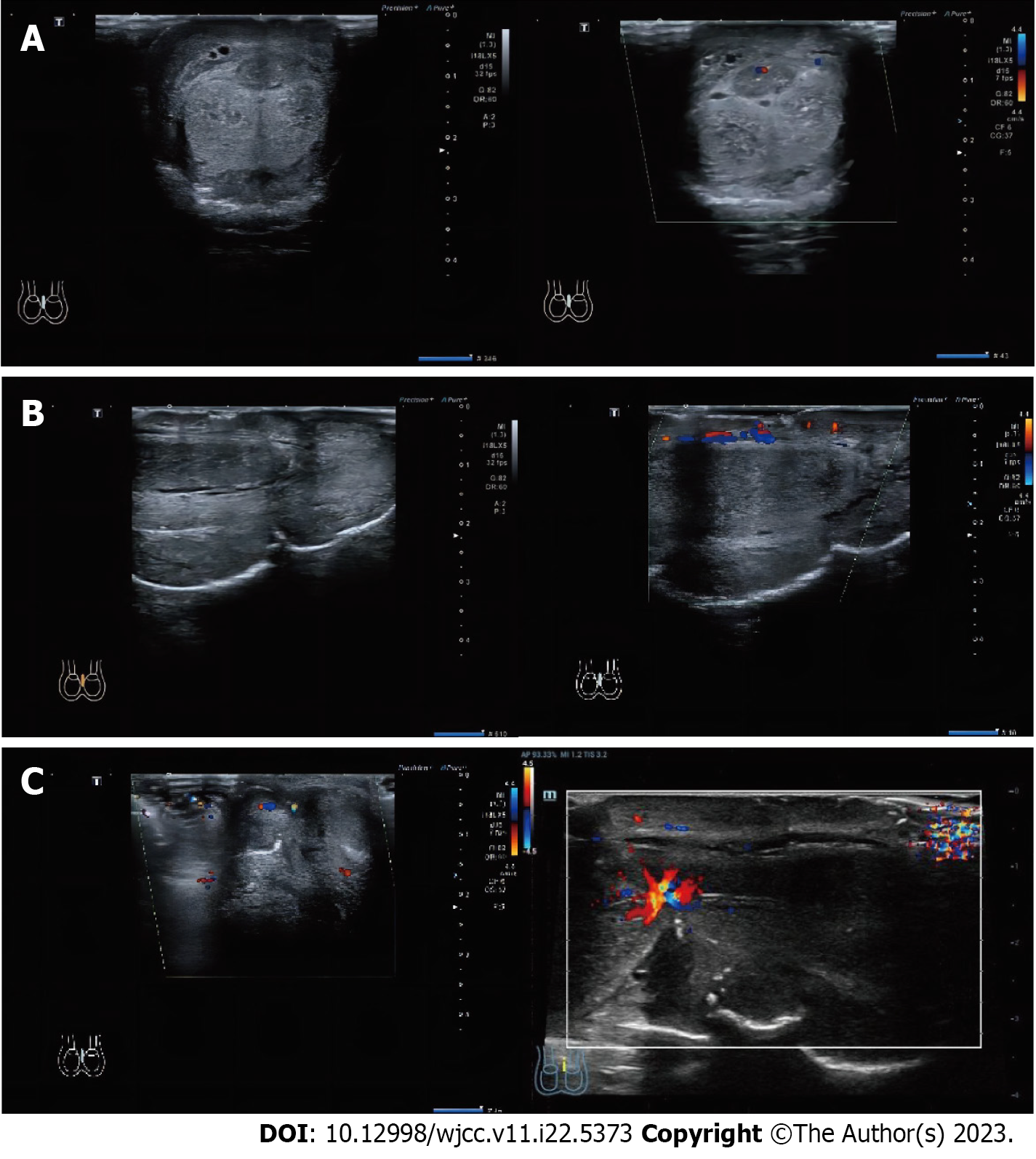
Figure 3 Preoperative Doppler ultrasonography.
The image depicts the color Doppler ultrasound display of the blood flow signal in the coronal and sagittal planes, as well as the strangulation site of the penis. A: Coronal plane; B: Vertical plane; C: Strangulation site.
- Citation: Maimaitiming ABLT, Mulati YLSD, Apizi ART, Li XD. Self-strangulation induced penile partial amputation: A case report. World J Clin Cases 2023; 11(22): 5373-5381
- URL: https://www.wjgnet.com/2307-8960/full/v11/i22/5373.htm
- DOI: https://dx.doi.org/10.12998/wjcc.v11.i22.5373


