Copyright
©The Author(s) 2023.
World J Clin Cases. Aug 6, 2023; 11(22): 5309-5315
Published online Aug 6, 2023. doi: 10.12998/wjcc.v11.i22.5309
Published online Aug 6, 2023. doi: 10.12998/wjcc.v11.i22.5309
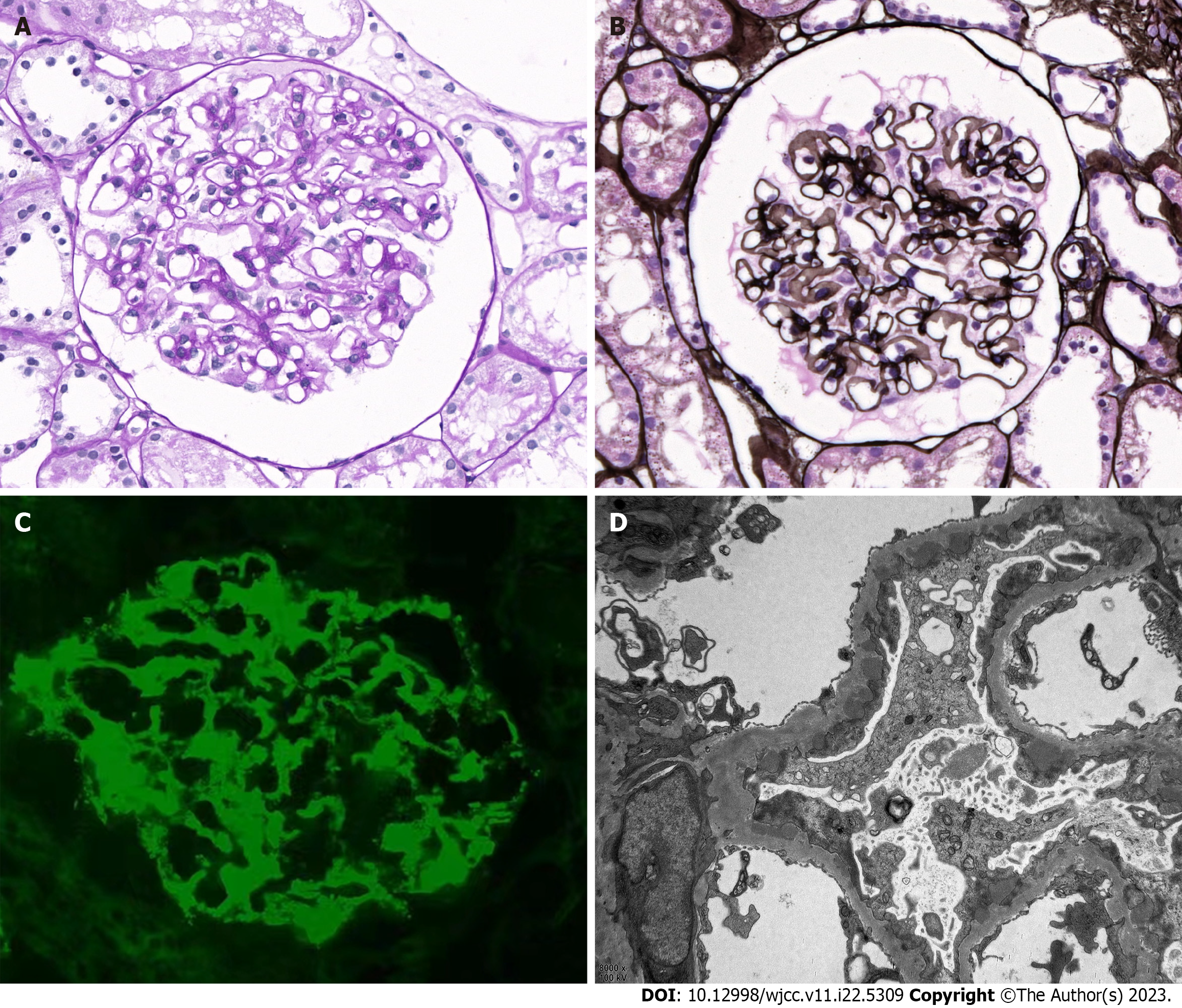
Figure 1 Biopsy findings in patients with human immunodeficiency virus infection and membranous nephropathy.
A: Histological analysis of biopsy tissues via light microscopy showed eosinophilic deposits under the epithelium and a thickening of the basement membrane with vacuoles and granules visualized by periodic acid-Schiff staining (× 400); B: Periodic acid methenamine silver staining visualization also highlights vacuoles and granules (× 400); C: Immunofluorescent evaluation of renal tissues (× 400) showed granular phospholipase A2 receptor-associated deposition along capillary loops; D: Electron microscopy showed subepithelial electron-dense deposits with the associated basement membrane, consistent with stage I-II membranous nephropathy (× 8000).
- Citation: Wang JL, Sun YL, Kang Z, Zhang SK, Yu CX, Zhang W, Xie H, Lin HL. Anti-phospholipase A2 receptor-associated membranous nephropathy with human immunodeficiency virus infection treated with telitacicept: A case report. World J Clin Cases 2023; 11(22): 5309-5315
- URL: https://www.wjgnet.com/2307-8960/full/v11/i22/5309.htm
- DOI: https://dx.doi.org/10.12998/wjcc.v11.i22.5309


