Copyright
©The Author(s) 2023.
World J Clin Cases. Jul 26, 2023; 11(21): 5122-5128
Published online Jul 26, 2023. doi: 10.12998/wjcc.v11.i21.5122
Published online Jul 26, 2023. doi: 10.12998/wjcc.v11.i21.5122
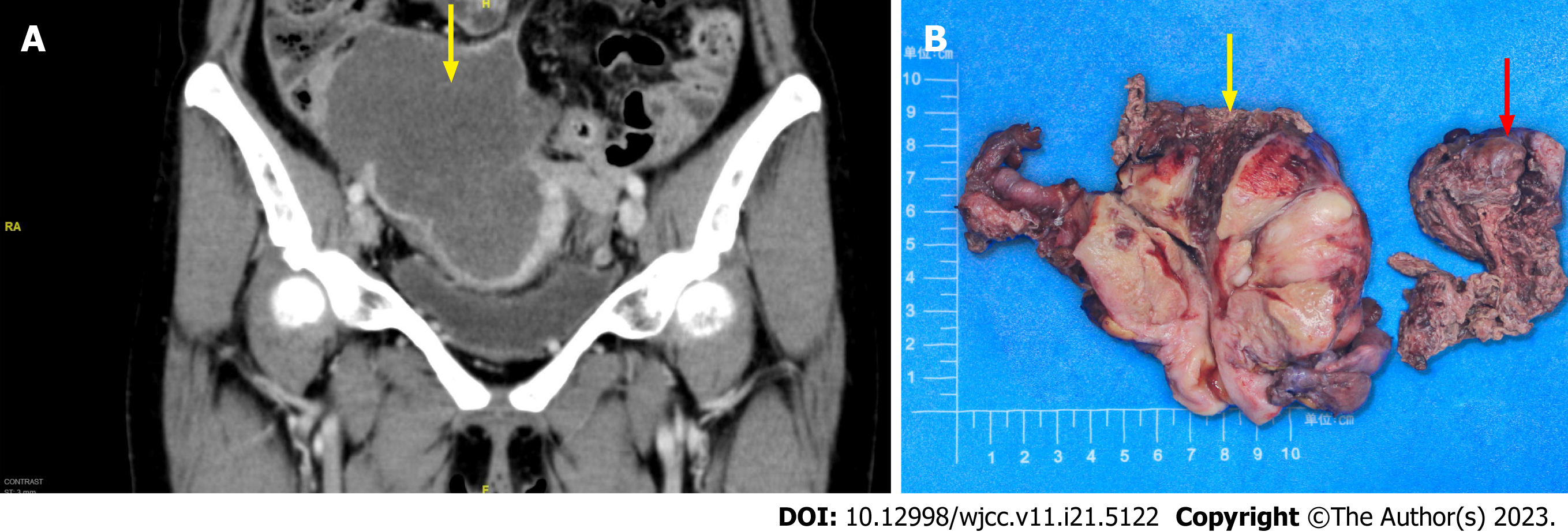
Figure 1 Computed tomography findings and macroscopic observation for case 1.
A: Computed tomography scan revealed a large solid mass in the right adnexal region, with a size of 12.8 cm × 11.6 cm × 7.8 cm (yellow arrow) leading to obstruction and dilatation of the middle and upper segment of the ureter, and invading the adjacent area; B: Right ovary was greatly enlarged and nodular with cystic and solid, reddish-white cut surface ureter (red arrow), and invaded the right lower segment of the ureter (yellow arrow).
- Citation: Zhou Y, Sun YW, Liu XY, Shen DH. Primary ovarian angiosarcoma: Two case reports and review of literature. World J Clin Cases 2023; 11(21): 5122-5128
- URL: https://www.wjgnet.com/2307-8960/full/v11/i21/5122.htm
- DOI: https://dx.doi.org/10.12998/wjcc.v11.i21.5122


