Copyright
©The Author(s) 2023.
World J Clin Cases. Jan 16, 2023; 11(2): 342-356
Published online Jan 16, 2023. doi: 10.12998/wjcc.v11.i2.342
Published online Jan 16, 2023. doi: 10.12998/wjcc.v11.i2.342
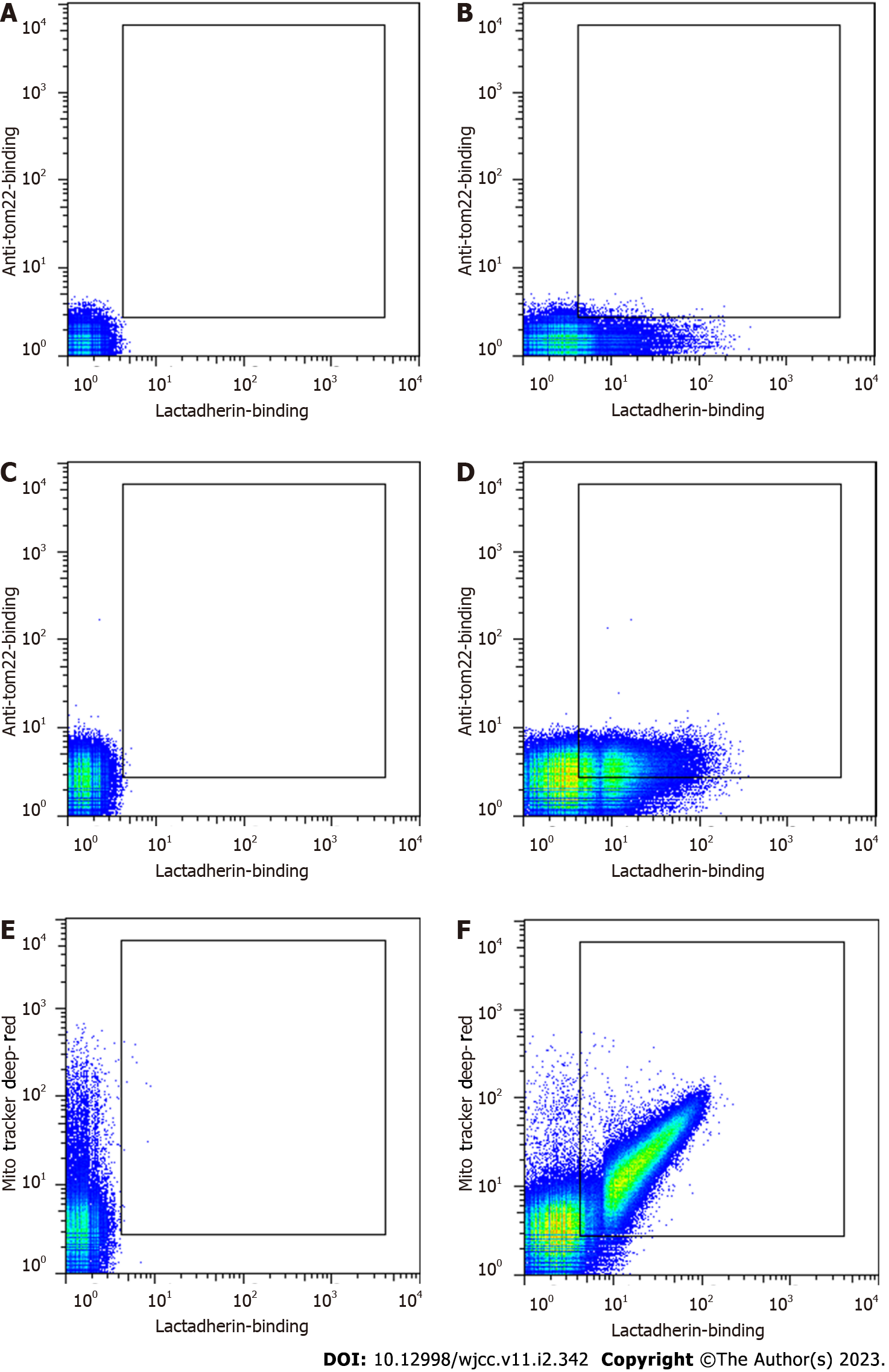
Figure 2 Representative dot plots of circulating microvesicles from a patient with sepsis.
Microvesicles (MVs) were labelled and analysed by flow cytometry. A: Unlabelled MVs (autofluorescence); B: MVs labelled with lactadherin-FITC; C: MVs labelled with anti-tom22-APC; D: MVs double stained with lactadherin-FITC and anti-tom22-APC; E: MVs labelled with MitoTracker Deep Red; F: MVs double stained with lactadherin-FITC and MitoTracker Deep Red.
- Citation: Zhang HJ, Li JY, Wang C, Zhong GQ. Microvesicles with mitochondrial content are increased in patients with sepsis and associated with inflammatory responses. World J Clin Cases 2023; 11(2): 342-356
- URL: https://www.wjgnet.com/2307-8960/full/v11/i2/342.htm
- DOI: https://dx.doi.org/10.12998/wjcc.v11.i2.342


