Copyright
©The Author(s) 2023.
World J Clin Cases. Jul 6, 2023; 11(19): 4698-4706
Published online Jul 6, 2023. doi: 10.12998/wjcc.v11.i19.4698
Published online Jul 6, 2023. doi: 10.12998/wjcc.v11.i19.4698
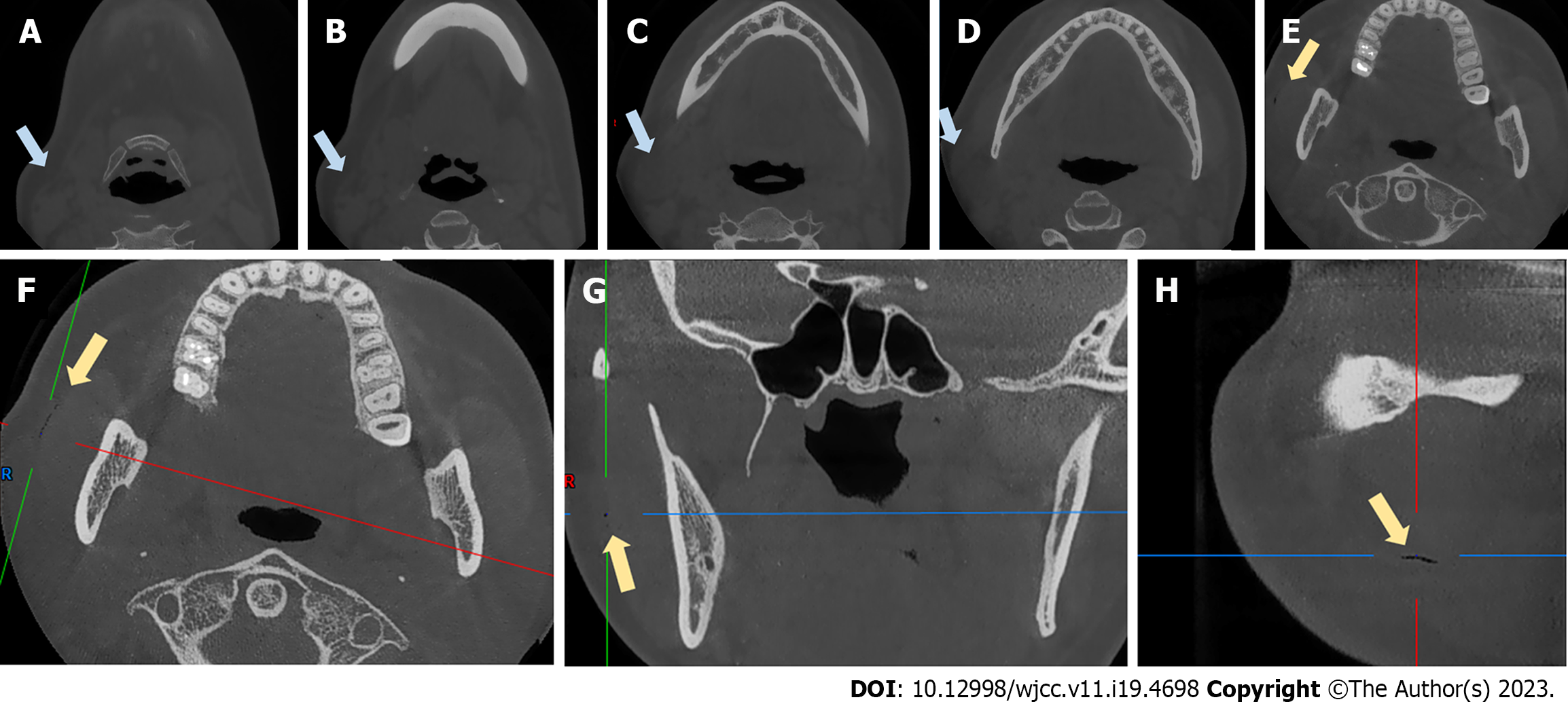
Figure 4 Cone-beam computed tomography scans obtained after emphysema developed.
A-E: Coronal views of the swelling; F-H: An air-filled fissure between the masseter muscle and soft tissue. Blue arrows: Swelling. Yellow arrows: The fissure.
- Citation: Bai YP, Sha JJ, Chai CC, Sun HP. With two episodes of right retromandibular angle subcutaneous emphysema during right upper molar crown preparation: A case report. World J Clin Cases 2023; 11(19): 4698-4706
- URL: https://www.wjgnet.com/2307-8960/full/v11/i19/4698.htm
- DOI: https://dx.doi.org/10.12998/wjcc.v11.i19.4698


