Copyright
©The Author(s) 2023.
World J Clin Cases. Jul 6, 2023; 11(19): 4648-4654
Published online Jul 6, 2023. doi: 10.12998/wjcc.v11.i19.4648
Published online Jul 6, 2023. doi: 10.12998/wjcc.v11.i19.4648
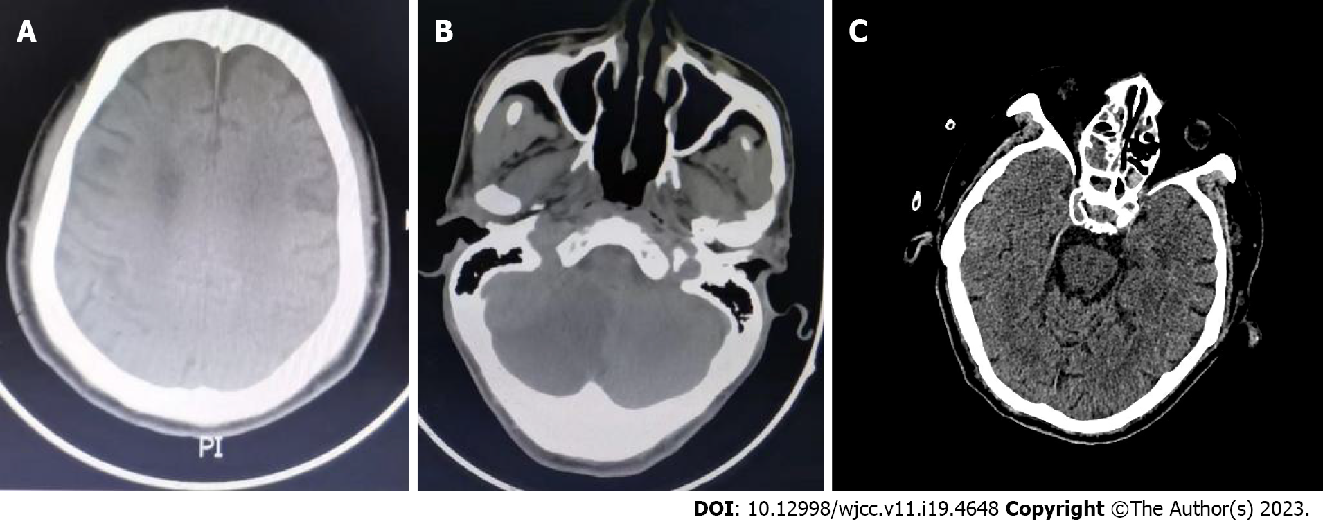
Figure 1 Computed tomography.
A: Case 1: Head computed tomography (CT) scan showed cerebral infarction in the bilateral parietal lobes with multiple small ischemic lesions; B: Case 2: The patient’s emergency head CT scan showed cerebral infarction in the left thalamus and right cerebellar hemisphere; C: Case 3: Head CT scan suggested the probability of lacunar infarction in the left brainstem.
- Citation: Yang L, Xu X, Wang L, Zeng KB, Wang XF. Edaravone administration and its potential association with a new clinical syndrome in cerebral infarction patients: Three case reports. World J Clin Cases 2023; 11(19): 4648-4654
- URL: https://www.wjgnet.com/2307-8960/full/v11/i19/4648.htm
- DOI: https://dx.doi.org/10.12998/wjcc.v11.i19.4648


