Copyright
©The Author(s) 2023.
World J Clin Cases. Jun 26, 2023; 11(18): 4446-4453
Published online Jun 26, 2023. doi: 10.12998/wjcc.v11.i18.4446
Published online Jun 26, 2023. doi: 10.12998/wjcc.v11.i18.4446
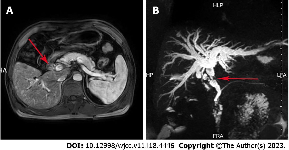
Figure 1 Imaging findings of the bile duct neoplasms.
A: Magnetic resonance imaging (MRI) of the abdomen and magnetic resonance cholang
- Citation: Jiao X, Zhai MM, Xing FZ, Wang XL. Simultaneously metastatic cholangiocarcinoma and small intestine cancer from breast cancer misdiagnosed as primary cholangiocarcinoma: A case report. World J Clin Cases 2023; 11(18): 4446-4453
- URL: https://www.wjgnet.com/2307-8960/full/v11/i18/4446.htm
- DOI: https://dx.doi.org/10.12998/wjcc.v11.i18.4446


