Copyright
©The Author(s) 2023.
World J Clin Cases. Jun 26, 2023; 11(18): 4360-4367
Published online Jun 26, 2023. doi: 10.12998/wjcc.v11.i18.4360
Published online Jun 26, 2023. doi: 10.12998/wjcc.v11.i18.4360
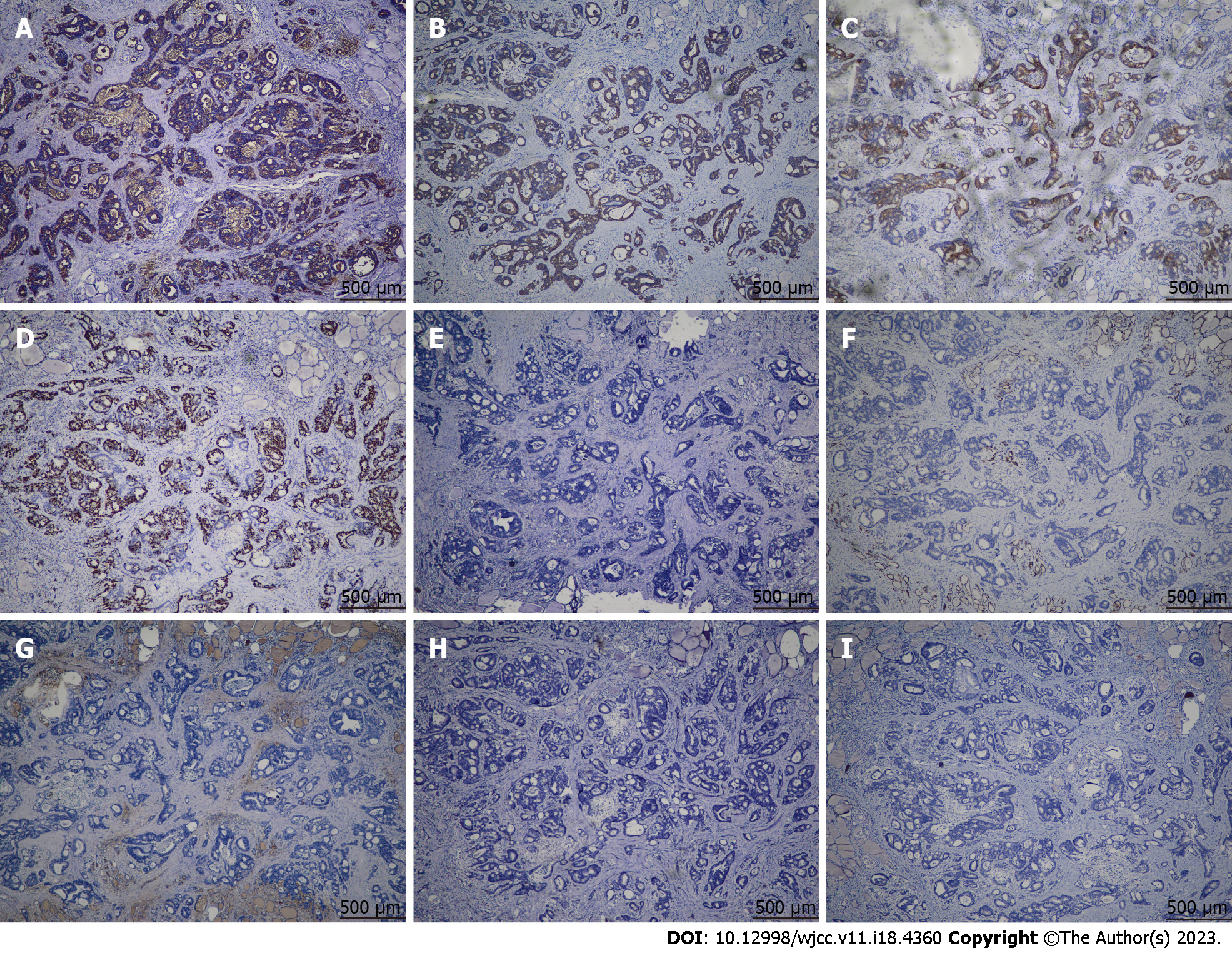
Figure 4 Immunohistochemical images showing metastatic carcinoma in the resected thyroid gland.
A-D: Tumor cells are positive for carcinoembryonic antigen (A), cytokeratin 20 (B), EMA (C), and Ki-67 (about 50%) (D); E-I: Tumor cells are negative for HBME1 (E), cytokeratin 7 (F), thyroglobulin (G), synaptophysin (H), and calcitonin (I).
- Citation: Chen Y, Kang QS, Zheng Y, Li FB. Solitary thyroid gland metastasis from rectal cancer: A case report and review of the literature. World J Clin Cases 2023; 11(18): 4360-4367
- URL: https://www.wjgnet.com/2307-8960/full/v11/i18/4360.htm
- DOI: https://dx.doi.org/10.12998/wjcc.v11.i18.4360


