Copyright
©The Author(s) 2023.
World J Clin Cases. Jun 6, 2023; 11(16): 3921-3928
Published online Jun 6, 2023. doi: 10.12998/wjcc.v11.i16.3921
Published online Jun 6, 2023. doi: 10.12998/wjcc.v11.i16.3921
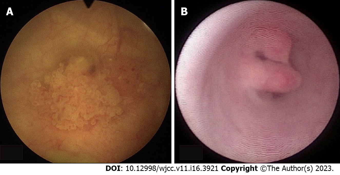
Figure 3 Photograph from ureteroscopy.
A: Photograph from ureteroscopy; cauliflower-like lesions over right posterior and right lateral wall junction of the bladder are observed; B: Photograph of the patient’s left ureter via ureteroscopy; an intraluminal bulging tumor located at the middle third of the ureteral cavity (approximately 0.8 cm) is observed. Histological examination of the lesion confirms the diagnosis of urothelial carcinoma.
- Citation: Tsai YC, Li CC, Chen BT, Wang CY. Coexistence of urinary tuberculosis and urothelial carcinoma: A case report. World J Clin Cases 2023; 11(16): 3921-3928
- URL: https://www.wjgnet.com/2307-8960/full/v11/i16/3921.htm
- DOI: https://dx.doi.org/10.12998/wjcc.v11.i16.3921


