Copyright
©The Author(s) 2023.
World J Clin Cases. May 26, 2023; 11(15): 3643-3650
Published online May 26, 2023. doi: 10.12998/wjcc.v11.i15.3643
Published online May 26, 2023. doi: 10.12998/wjcc.v11.i15.3643
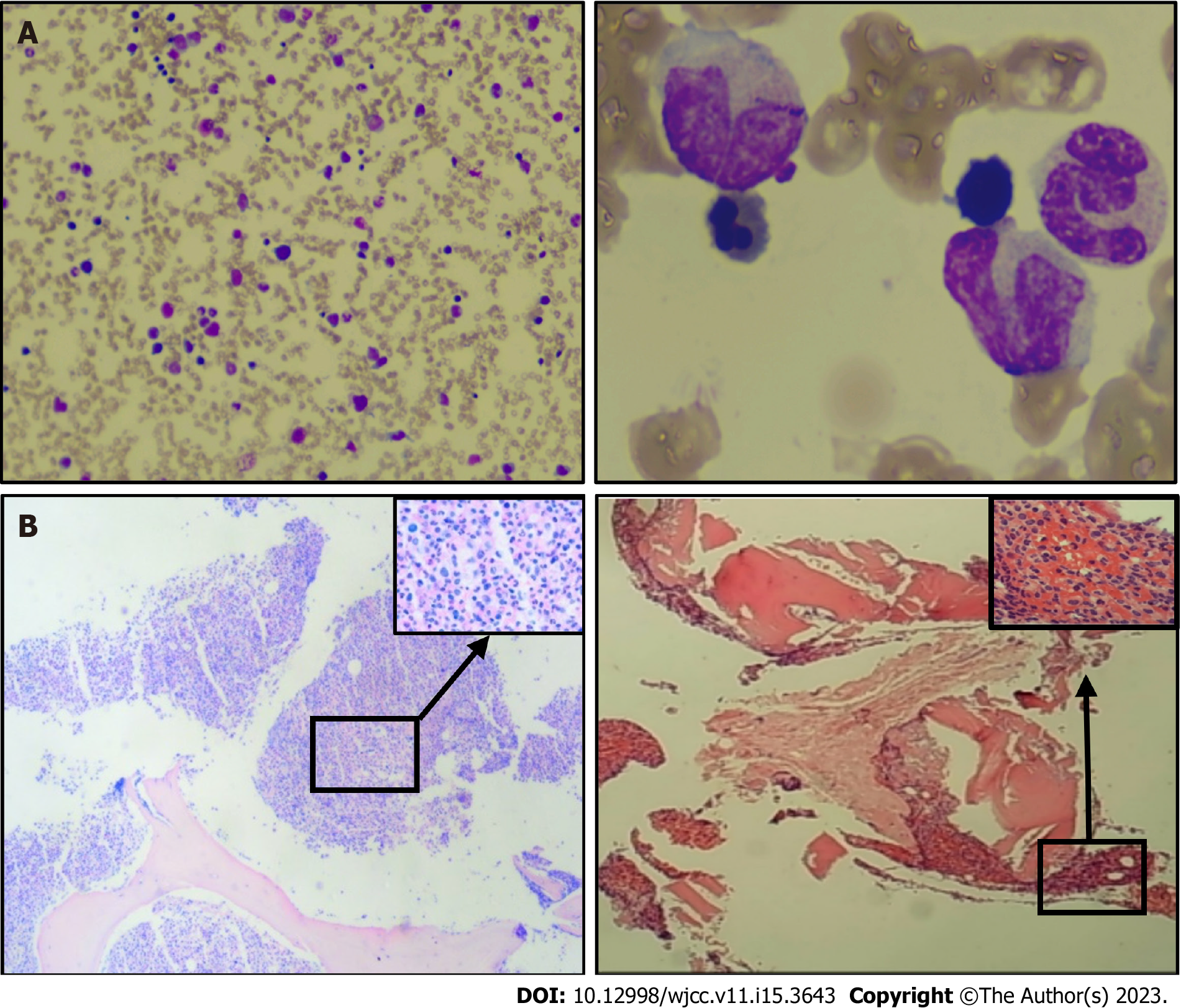
Figure 1 Morphological and biopsy results of chronic myelomonocytic leukemia bone marrow analysis.
A: Pelger deformed granules (100× magnification), exhibiting abnormal proliferation of monocytes and occasional naïve monocytes (with well-differentiated monocytes accounting for 45% of total monocytes), inhibited erythroid hyperplasia, three megakaryocytes, and rare platelet; B: Increased naïve-stage cells, with an increased granulocyte-to-nucleated red blood cell ratio. The granular lineage predominantly comprised intermediate (lower-stage) and visible monocytes, based on reticulocyte staining (MF-0 grade; ×4 and ×40 magnification).
- Citation: Deng LJ, Dong Y, Li MM, Sun CG. Co-existing squamous cell carcinoma and chronic myelomonocytic leukemia with ASXL1 and EZH2 gene mutations: A case report. World J Clin Cases 2023; 11(15): 3643-3650
- URL: https://www.wjgnet.com/2307-8960/full/v11/i15/3643.htm
- DOI: https://dx.doi.org/10.12998/wjcc.v11.i15.3643


