Copyright
©The Author(s) 2023.
World J Clin Cases. May 26, 2023; 11(15): 3522-3532
Published online May 26, 2023. doi: 10.12998/wjcc.v11.i15.3522
Published online May 26, 2023. doi: 10.12998/wjcc.v11.i15.3522
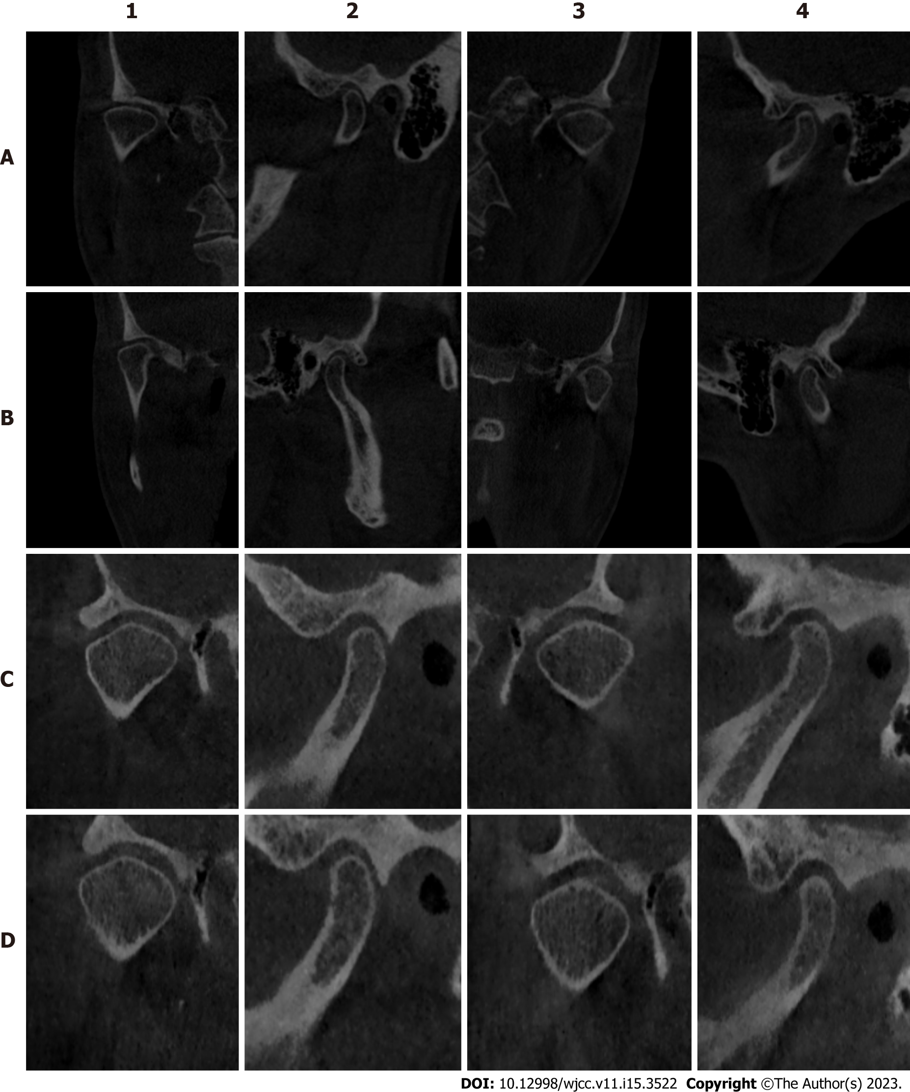
Figure 2 Cone beam computed tomography imaging.
A: Cone beam computed tomography (CBCT) at the beginning; B: CBCT after wearing an occlusal pad for 3 mo; C: CBCT 1 mo after wearing the temporary crown; D: CBCT rechecked 2 wk after wearing the final denture. 1 and 2 are the coronal and sagittal planes of the right temporomandibular joint, respectively; 3 and 4 are the coronal and sagittal planes of the left temporomandibular joint, respectively.
- Citation: Hou C, Zhu HZ, Xue B, Song HJ, Yang YB, Wang XX, Sun HQ. New clinical application of digital intraoral scanning technology in occlusal reconstruction: A case report. World J Clin Cases 2023; 11(15): 3522-3532
- URL: https://www.wjgnet.com/2307-8960/full/v11/i15/3522.htm
- DOI: https://dx.doi.org/10.12998/wjcc.v11.i15.3522


