Copyright
©The Author(s) 2023.
World J Clin Cases. May 16, 2023; 11(14): 3362-3368
Published online May 16, 2023. doi: 10.12998/wjcc.v11.i14.3362
Published online May 16, 2023. doi: 10.12998/wjcc.v11.i14.3362
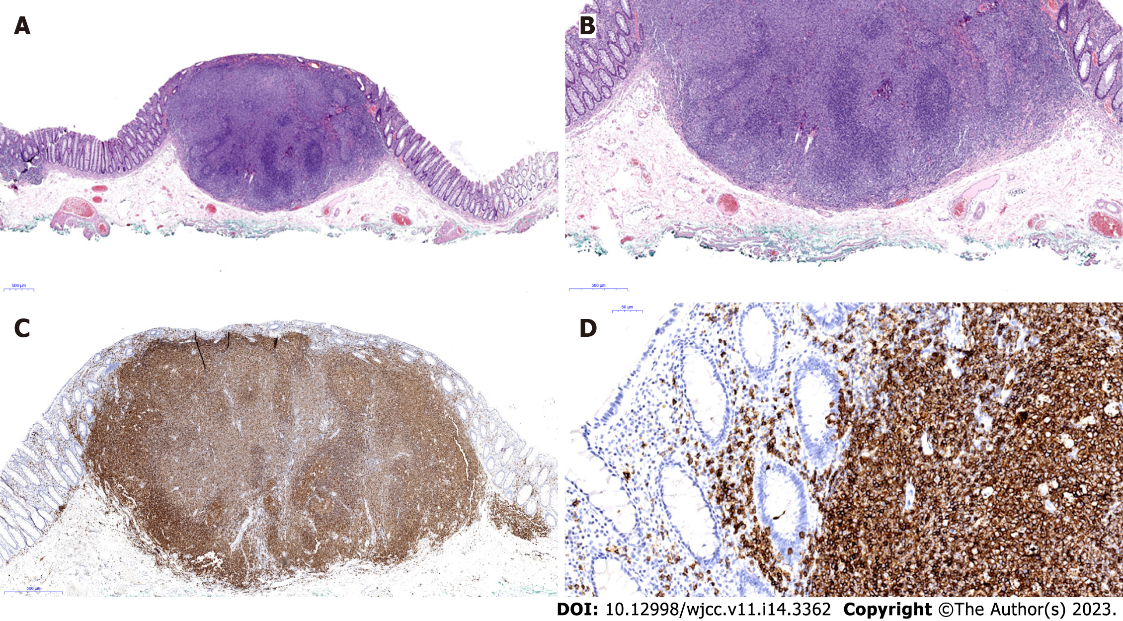
Figure 4 Microscopic findings.
A: Endoscopic biopsy specimens show a dense aggregate of lymphoma cells in the lamina propria (hematoxylin and eosin staining, ×20); B: Lymphoma cells infiltrate the mucosal layer above the subepithelial layer (hematoxylin and eosin staining, ×40); C: Immunohistochemical staining shows aggregate of lymphoma cells that stained positive for the B-cell marker CD20 (×400); D: Characteristic lymphocyte-epithelial lesion of CD20-positive lymphoma cells was also observed (x400).
- Citation: Lee WS, Noh MG, Joo YE. Primary rectal mucosa-associated lymphoid tissue lymphoma treated with only endoscopic submucosal dissection: A case report. World J Clin Cases 2023; 11(14): 3362-3368
- URL: https://www.wjgnet.com/2307-8960/full/v11/i14/3362.htm
- DOI: https://dx.doi.org/10.12998/wjcc.v11.i14.3362


