Copyright
©The Author(s) 2023.
World J Clin Cases. May 16, 2023; 11(14): 3295-3303
Published online May 16, 2023. doi: 10.12998/wjcc.v11.i14.3295
Published online May 16, 2023. doi: 10.12998/wjcc.v11.i14.3295
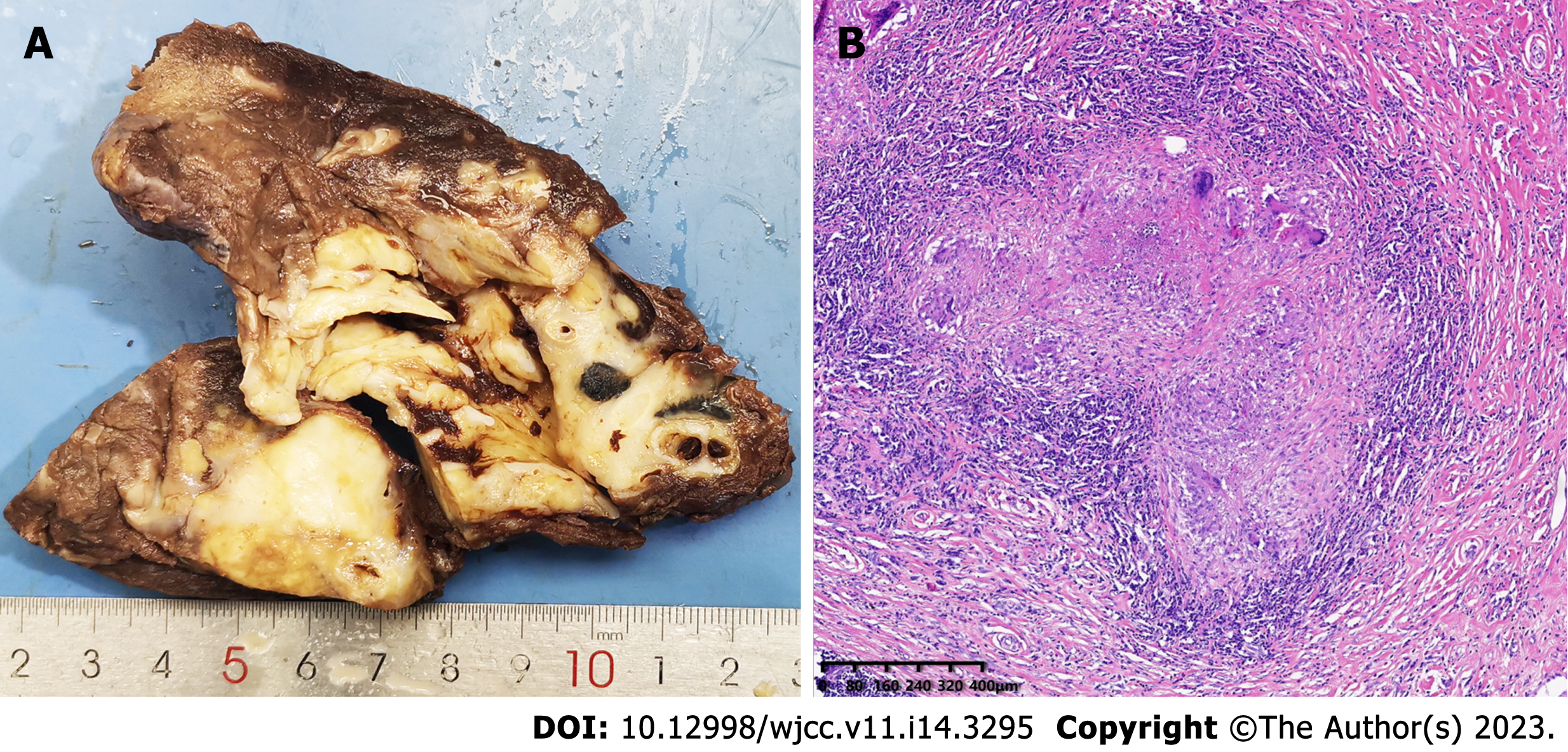
Figure 4 Macroscopic and microscopic features of the left lower lobe mass.
A: The mass was firm, approximately 6 cm in diameter, and pale in color when sectioned; and B: Hematoxylin and eosin stained section revealed that the left lower lobe mass was an inflammatory granuloma. Interstitial fibrous tissue hyperplasia, inflammatory cell infiltration, and multinucleated giant cell reaction were all observed under the microscope.
- Citation: Guo XZ, Gong LH, Wang WX, Yang DS, Zhang BH, Zhou ZT, Yu XH. Chronic pulmonary mucormycosis caused by rhizopus microsporus mimics lung carcinoma in an immunocompetent adult: A case report. World J Clin Cases 2023; 11(14): 3295-3303
- URL: https://www.wjgnet.com/2307-8960/full/v11/i14/3295.htm
- DOI: https://dx.doi.org/10.12998/wjcc.v11.i14.3295


