Copyright
©The Author(s) 2023.
World J Clin Cases. May 16, 2023; 11(14): 3295-3303
Published online May 16, 2023. doi: 10.12998/wjcc.v11.i14.3295
Published online May 16, 2023. doi: 10.12998/wjcc.v11.i14.3295
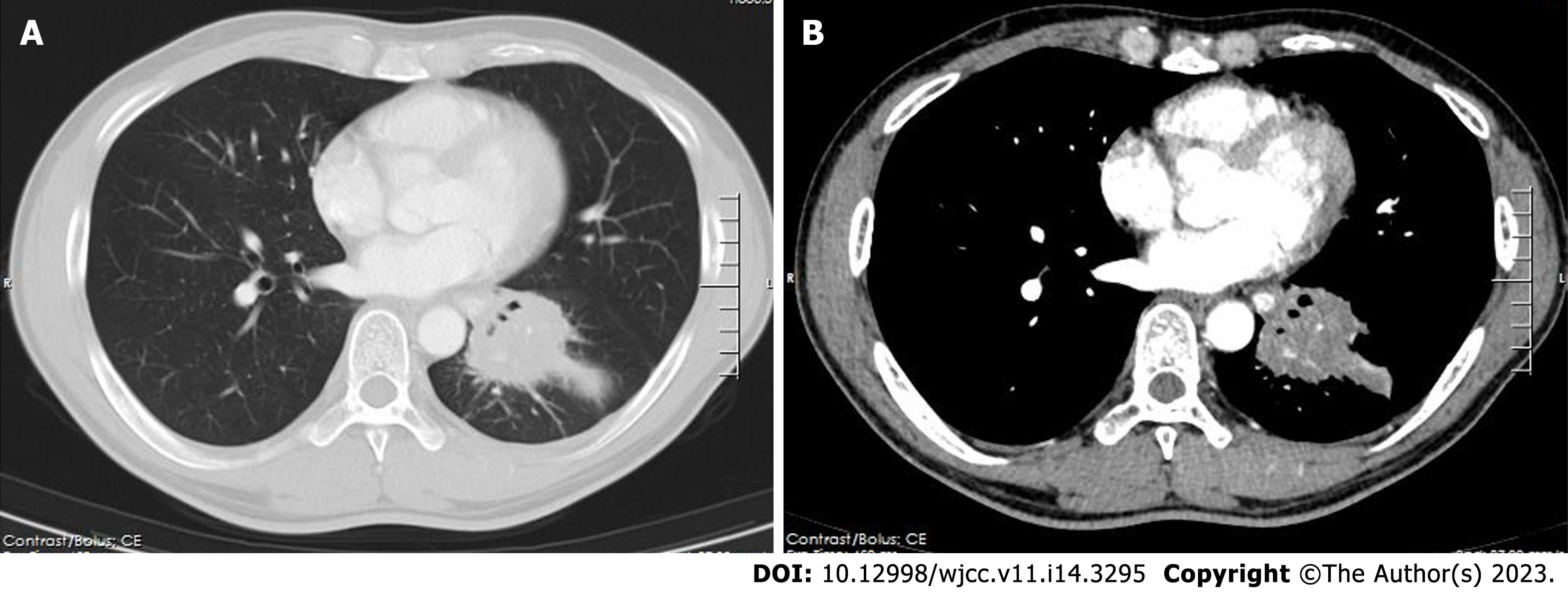
Figure 1 High-resolution chest contrast-enhanced computed tomography.
A: Lung window; and B: Mediastinal window. A mass in the left lower lobe with a diameter of approximately 6 cm. It was slightly enhanced and mainly located in the lateral segment (S9) and posterior segment (S10). The pulmonary vasculature was still looming in this mass, but there was bronchial occlusion of the affected lung segments.
- Citation: Guo XZ, Gong LH, Wang WX, Yang DS, Zhang BH, Zhou ZT, Yu XH. Chronic pulmonary mucormycosis caused by rhizopus microsporus mimics lung carcinoma in an immunocompetent adult: A case report. World J Clin Cases 2023; 11(14): 3295-3303
- URL: https://www.wjgnet.com/2307-8960/full/v11/i14/3295.htm
- DOI: https://dx.doi.org/10.12998/wjcc.v11.i14.3295


